Cobalt »
PDB 8sfc-8xc3 »
8tct »
Cobalt in PDB 8tct: Structure of 3K-Glch Bound Bacteroides Thetaiotaomicron 3-Keto-Beta- Glucopyranoside-1,2-Lyase BT1
Protein crystallography data
The structure of Structure of 3K-Glch Bound Bacteroides Thetaiotaomicron 3-Keto-Beta- Glucopyranoside-1,2-Lyase BT1, PDB code: 8tct
was solved by
A.C.Lazarski,
L.J.Worrall,
N.C.J.Strynadka,
with X-Ray Crystallography technique. A brief refinement statistics is given in the table below:
| Resolution Low / High (Å) | 45.69 / 1.86 |
| Space group | P 32 2 1 |
| Cell size a, b, c (Å), α, β, γ (°) | 122.287, 122.287, 68.747, 90, 90, 120 |
| R / Rfree (%) | 16.8 / 18.3 |
Cobalt Binding Sites:
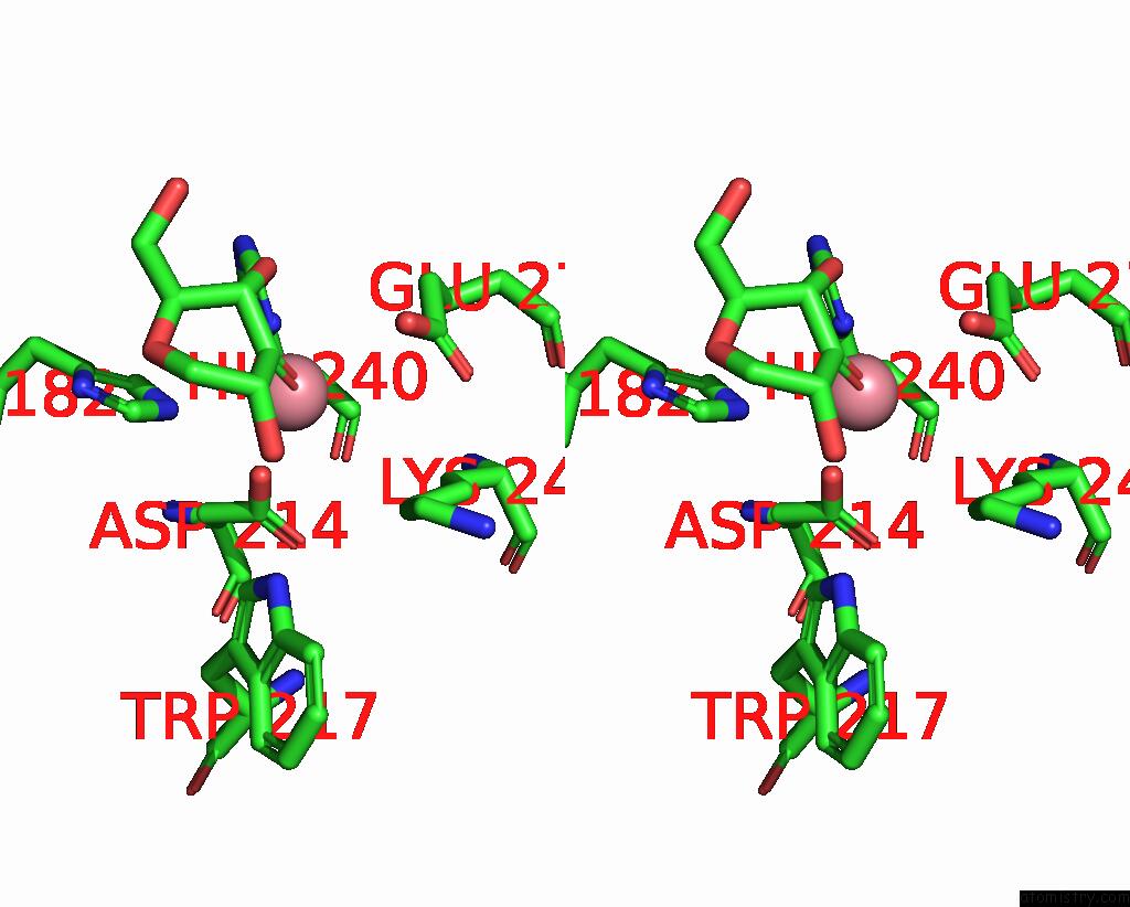
The binding sites of Cobalt atom in the Structure of 3K-Glch Bound Bacteroides Thetaiotaomicron 3-Keto-Beta- Glucopyranoside-1,2-Lyase BT1
(pdb code 8tct). This binding sites where shown within
5.0 Angstroms radius around Cobalt atom.
In total only one binding site of Cobalt was determined in the Structure of 3K-Glch Bound Bacteroides Thetaiotaomicron 3-Keto-Beta- Glucopyranoside-1,2-Lyase BT1, PDB code: 8tct:
In total only one binding site of Cobalt was determined in the Structure of 3K-Glch Bound Bacteroides Thetaiotaomicron 3-Keto-Beta- Glucopyranoside-1,2-Lyase BT1, PDB code: 8tct:
Cobalt binding site 1 out of 1 in 8tct
Go back to
Cobalt binding site 1 out
of 1 in the Structure of 3K-Glch Bound Bacteroides Thetaiotaomicron 3-Keto-Beta- Glucopyranoside-1,2-Lyase BT1
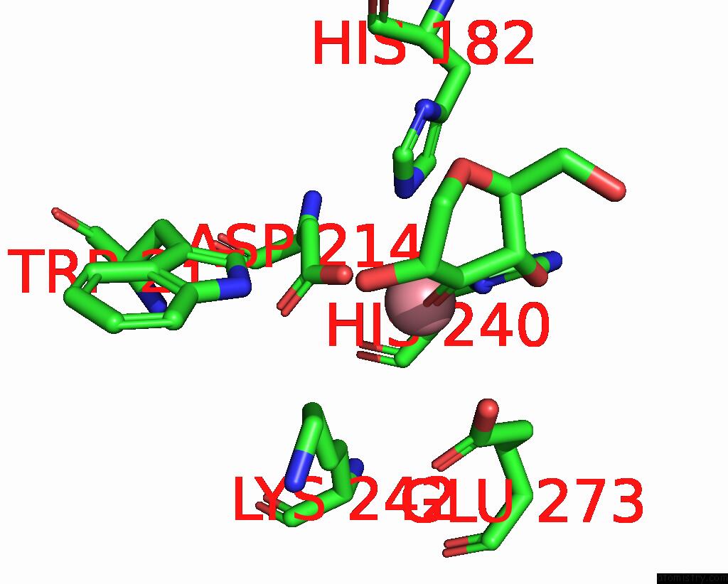
Mono view
Stereo pair view
Mono view
Stereo pair view
A full contact list of Cobalt with other atoms in the Co binding
site number 1 of Structure of 3K-Glch Bound Bacteroides Thetaiotaomicron 3-Keto-Beta- Glucopyranoside-1,2-Lyase BT1 within 5.0Å range:
|
Reference:
S.A.Nasseri,
A.C.Lazarski,
I.L.Lemmer,
C.Y.Zhang,
E.Brencher,
H.Chen,
L.Sim,
D.Panwar,
L.Betschart,
L.J.Worrall,
H.Brumer,
N.C.J.Strynadka,
S.G.Withers.
An Alternative Broad-Specificity Pathway For Glycan Breakdown in Bacteria Nature 2024.
ISSN: ESSN 1476-4687
Page generated: Tue Jul 30 20:03:19 2024
ISSN: ESSN 1476-4687
Last articles
Zn in 9MJ5Zn in 9HNW
Zn in 9G0L
Zn in 9FNE
Zn in 9DZN
Zn in 9E0I
Zn in 9D32
Zn in 9DAK
Zn in 8ZXC
Zn in 8ZUF