Cobalt »
PDB 8xhj-9icb »
9grd »
Cobalt in PDB 9grd: Cryo-Electron Microscopy Structure of Glucose/Xylose Isomerase From Streptomyces Rubiginosus with Cobalt Ions in the Active Site
Enzymatic activity of Cryo-Electron Microscopy Structure of Glucose/Xylose Isomerase From Streptomyces Rubiginosus with Cobalt Ions in the Active Site
All present enzymatic activity of Cryo-Electron Microscopy Structure of Glucose/Xylose Isomerase From Streptomyces Rubiginosus with Cobalt Ions in the Active Site:
5.3.1.5;
5.3.1.5;
Cobalt Binding Sites:
The binding sites of Cobalt atom in the Cryo-Electron Microscopy Structure of Glucose/Xylose Isomerase From Streptomyces Rubiginosus with Cobalt Ions in the Active Site
(pdb code 9grd). This binding sites where shown within
5.0 Angstroms radius around Cobalt atom.
In total 8 binding sites of Cobalt where determined in the Cryo-Electron Microscopy Structure of Glucose/Xylose Isomerase From Streptomyces Rubiginosus with Cobalt Ions in the Active Site, PDB code: 9grd:
Jump to Cobalt binding site number: 1; 2; 3; 4; 5; 6; 7; 8;
In total 8 binding sites of Cobalt where determined in the Cryo-Electron Microscopy Structure of Glucose/Xylose Isomerase From Streptomyces Rubiginosus with Cobalt Ions in the Active Site, PDB code: 9grd:
Jump to Cobalt binding site number: 1; 2; 3; 4; 5; 6; 7; 8;
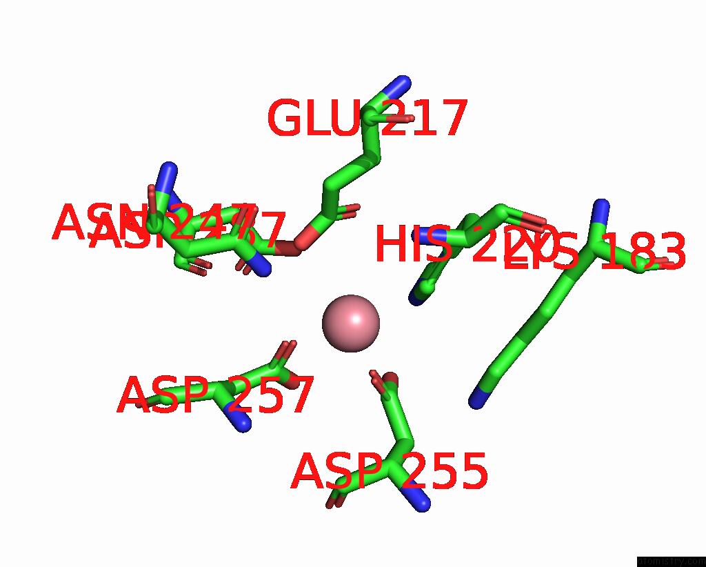
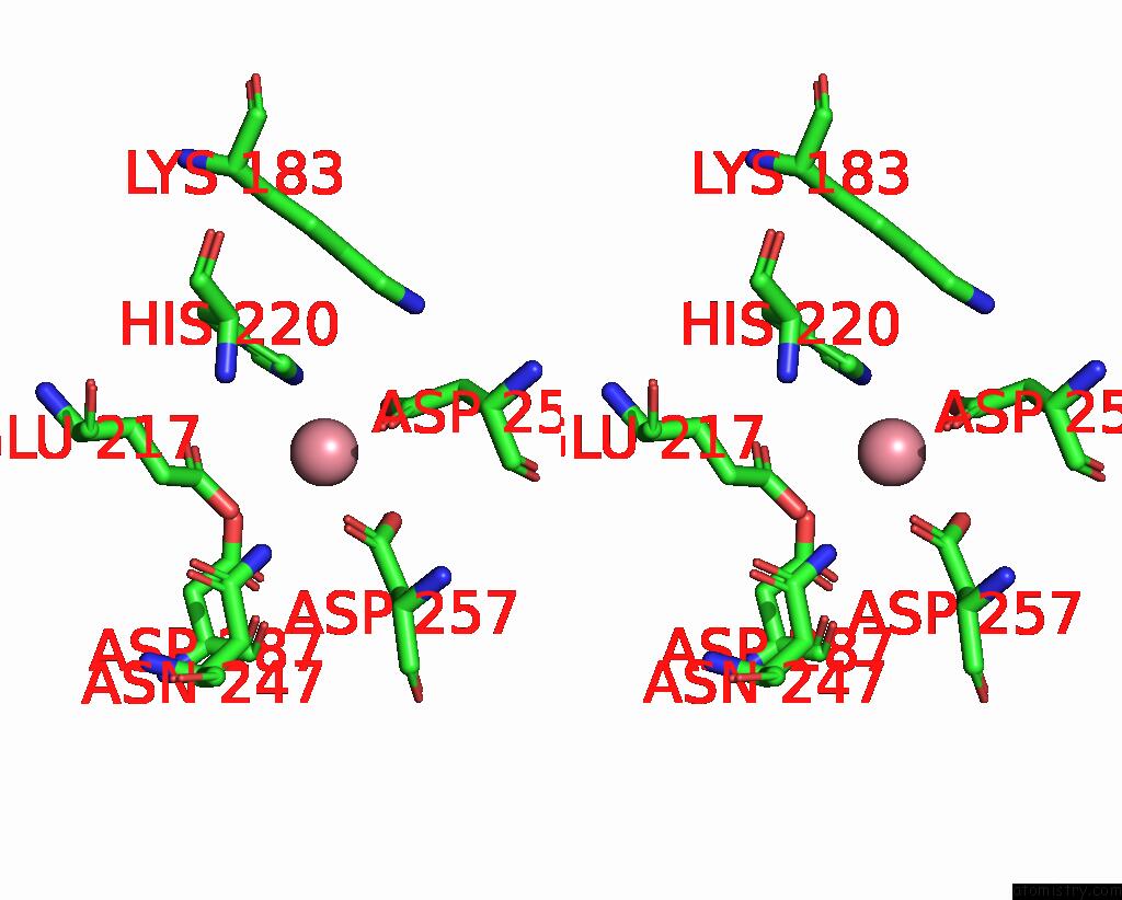
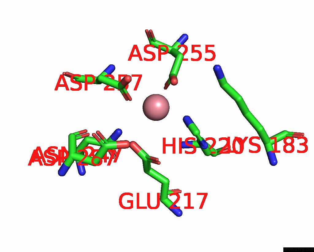
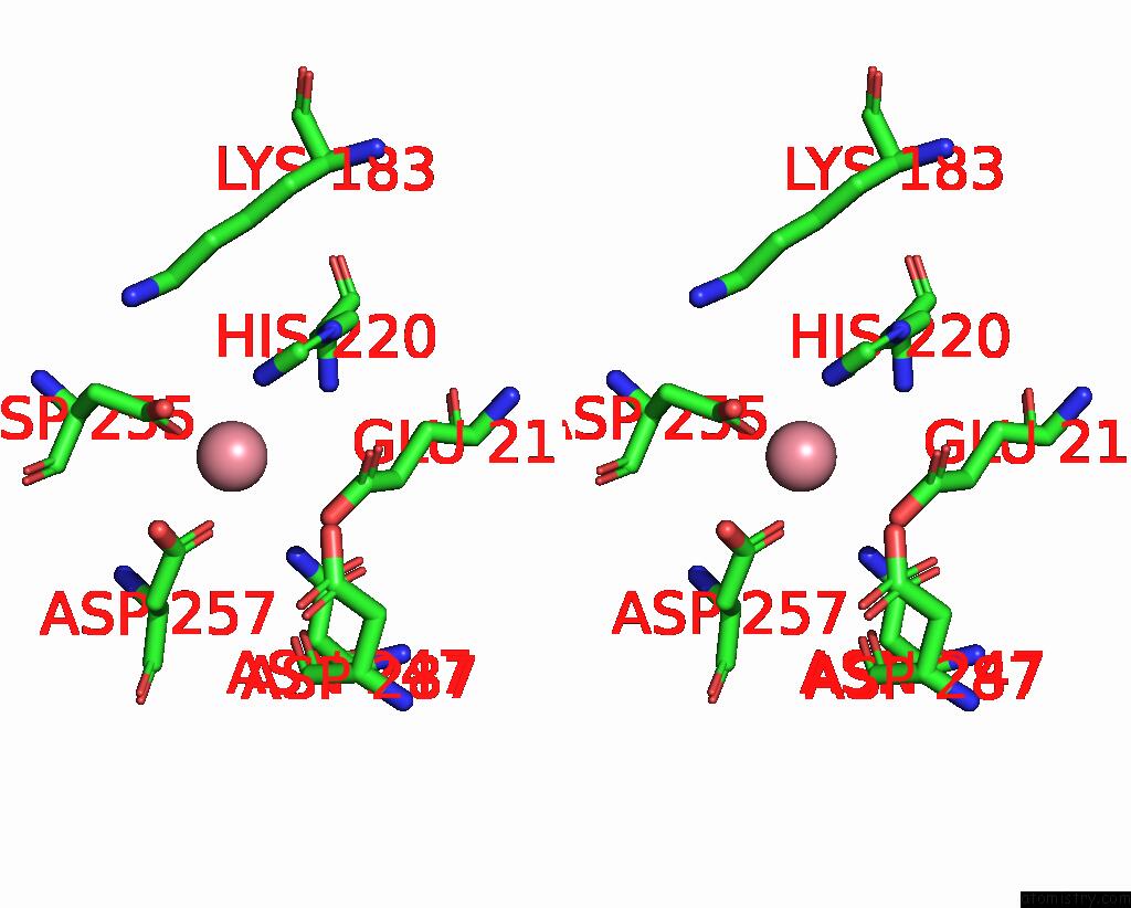
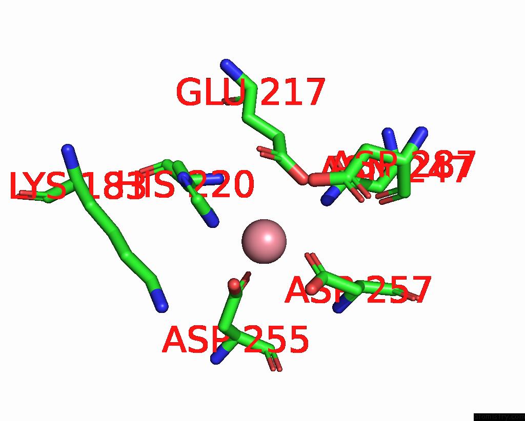
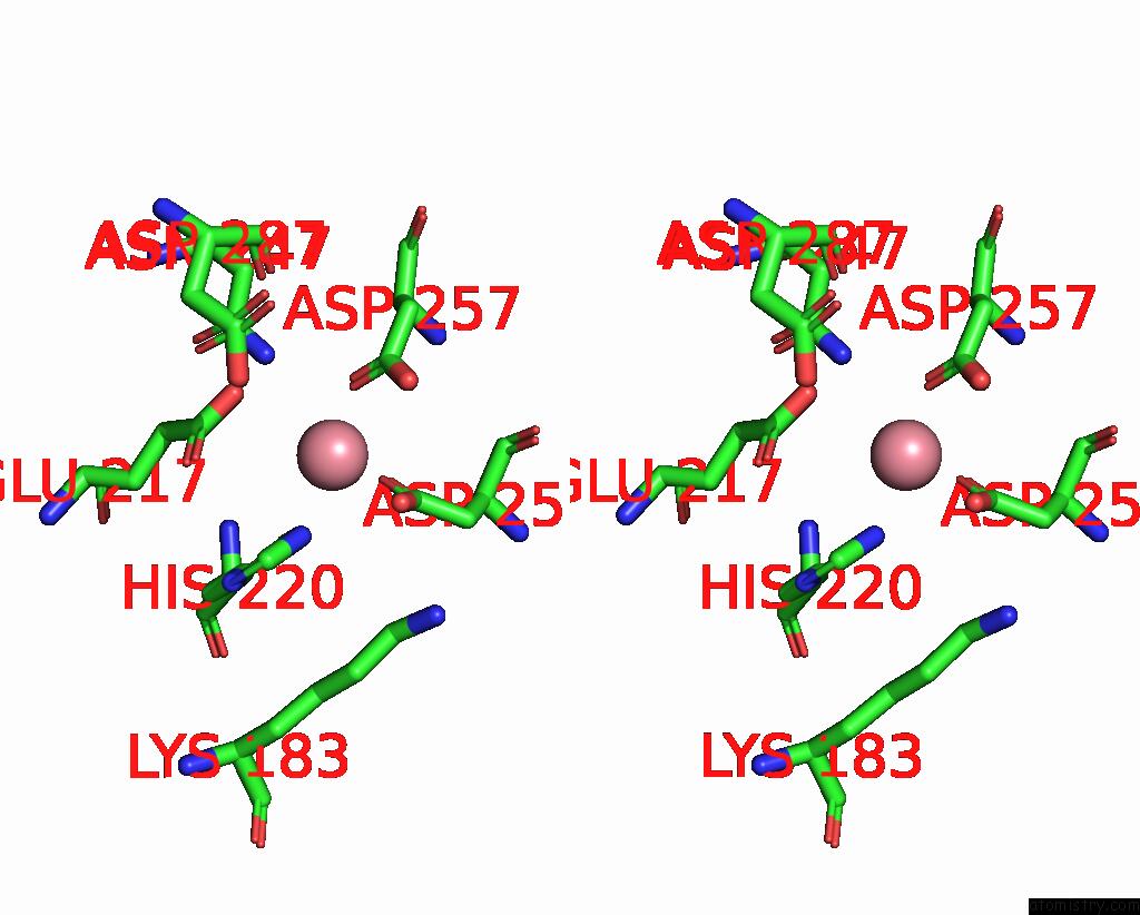
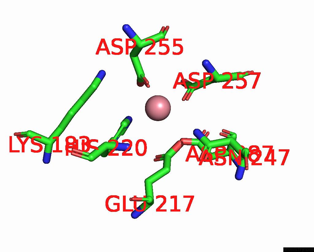
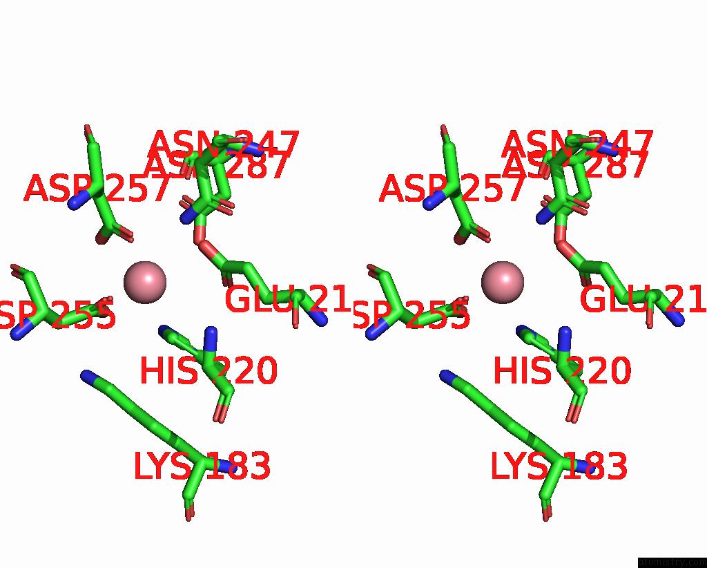
Cobalt binding site 1 out of 8 in 9grd
Go back to
Cobalt binding site 1 out
of 8 in the Cryo-Electron Microscopy Structure of Glucose/Xylose Isomerase From Streptomyces Rubiginosus with Cobalt Ions in the Active Site
Mono view
Stereo pair view
Mono view
Stereo pair view
A full contact list of Cobalt with other atoms in the Co binding
site number 1 of Cryo-Electron Microscopy Structure of Glucose/Xylose Isomerase From Streptomyces Rubiginosus with Cobalt Ions in the Active Site within 5.0Å range:
|
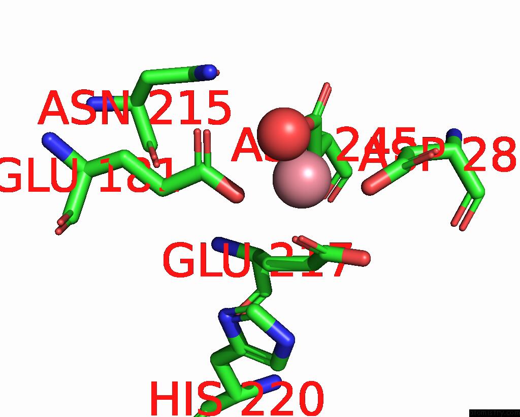
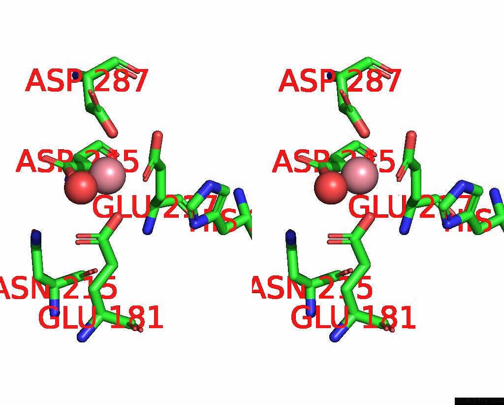
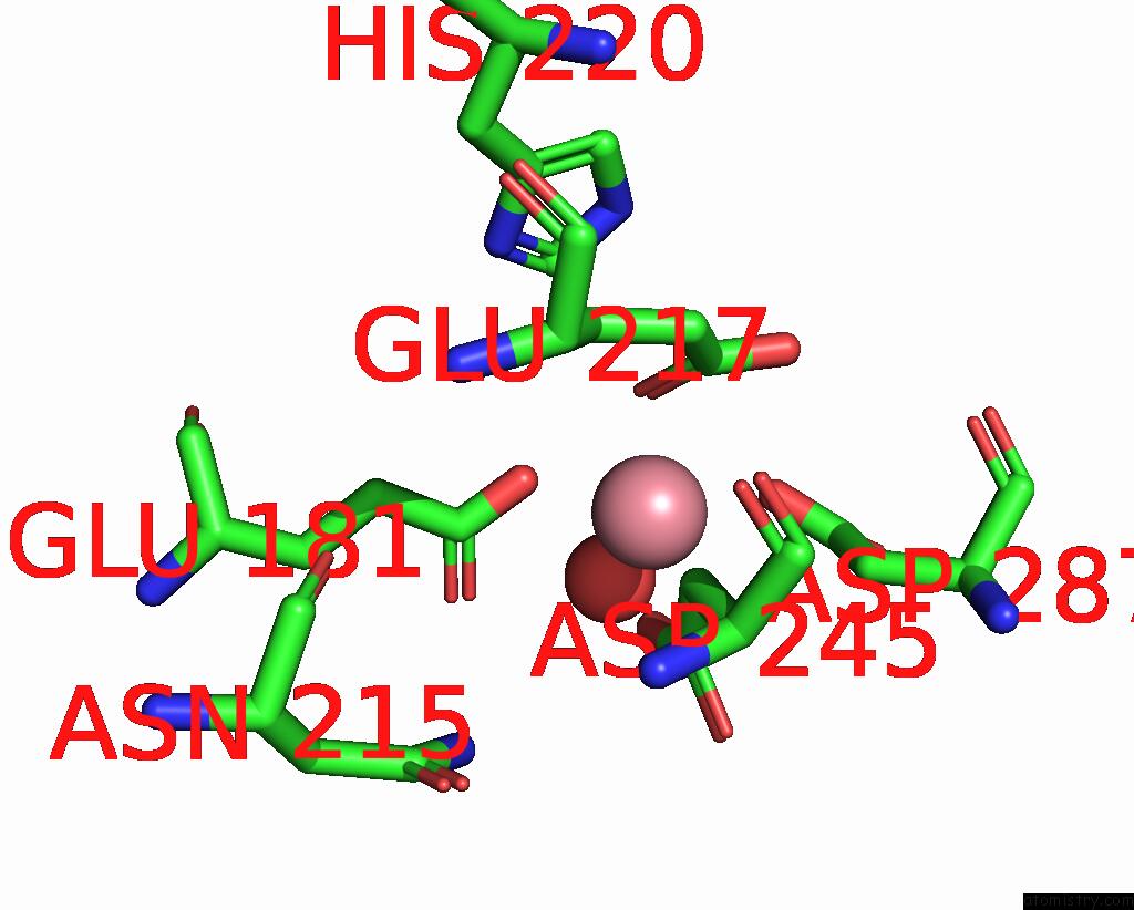
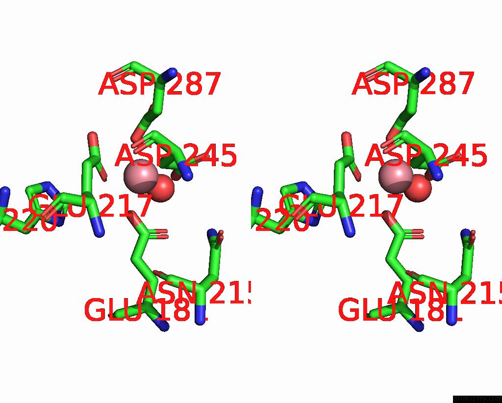
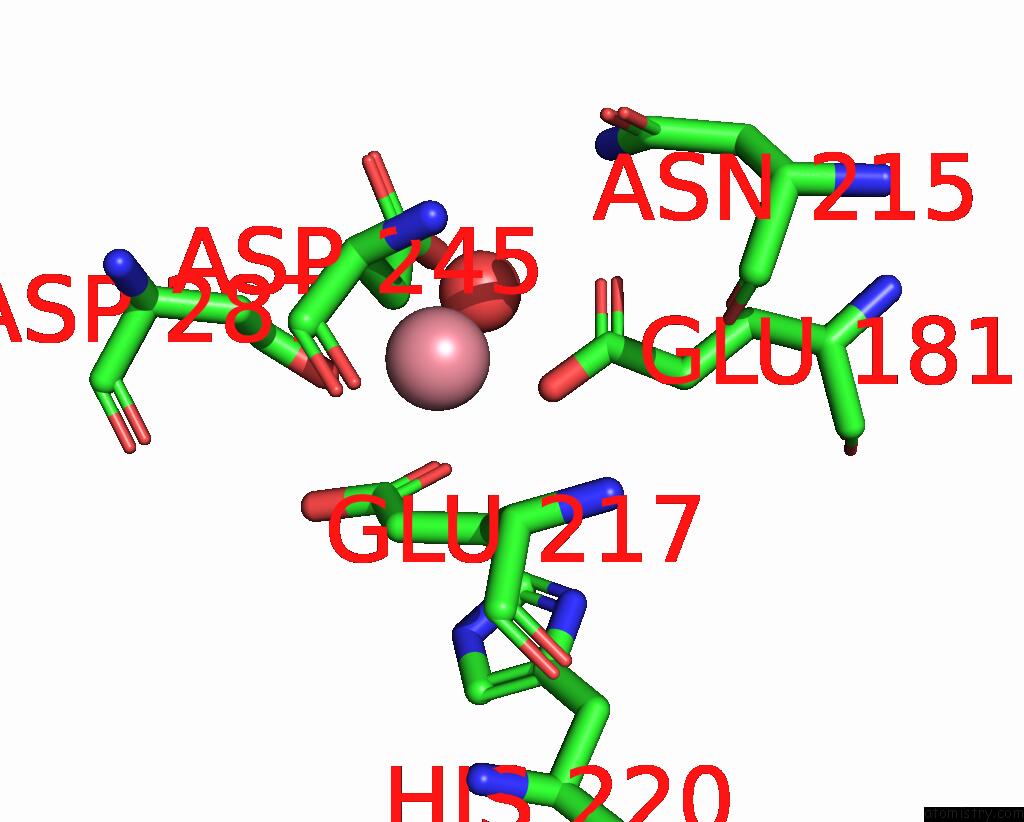
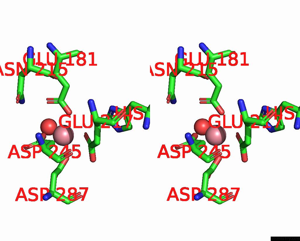
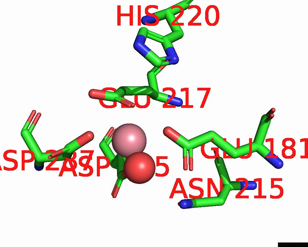
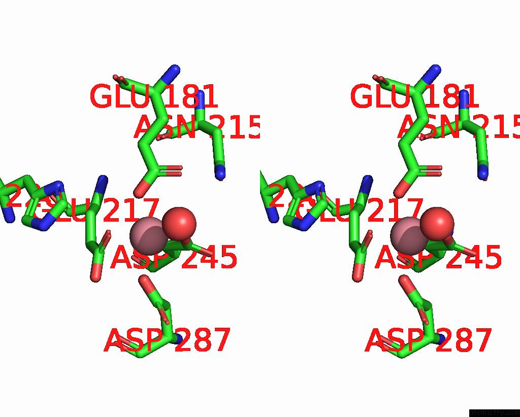
Cobalt binding site 2 out of 8 in 9grd
Go back to
Cobalt binding site 2 out
of 8 in the Cryo-Electron Microscopy Structure of Glucose/Xylose Isomerase From Streptomyces Rubiginosus with Cobalt Ions in the Active Site
Mono view
Stereo pair view
Mono view
Stereo pair view
A full contact list of Cobalt with other atoms in the Co binding
site number 2 of Cryo-Electron Microscopy Structure of Glucose/Xylose Isomerase From Streptomyces Rubiginosus with Cobalt Ions in the Active Site within 5.0Å range:
|
Cobalt binding site 3 out of 8 in 9grd
Go back to
Cobalt binding site 3 out
of 8 in the Cryo-Electron Microscopy Structure of Glucose/Xylose Isomerase From Streptomyces Rubiginosus with Cobalt Ions in the Active Site
Mono view
Stereo pair view
Mono view
Stereo pair view
A full contact list of Cobalt with other atoms in the Co binding
site number 3 of Cryo-Electron Microscopy Structure of Glucose/Xylose Isomerase From Streptomyces Rubiginosus with Cobalt Ions in the Active Site within 5.0Å range:
|
Cobalt binding site 4 out of 8 in 9grd
Go back to
Cobalt binding site 4 out
of 8 in the Cryo-Electron Microscopy Structure of Glucose/Xylose Isomerase From Streptomyces Rubiginosus with Cobalt Ions in the Active Site
Mono view
Stereo pair view
Mono view
Stereo pair view
A full contact list of Cobalt with other atoms in the Co binding
site number 4 of Cryo-Electron Microscopy Structure of Glucose/Xylose Isomerase From Streptomyces Rubiginosus with Cobalt Ions in the Active Site within 5.0Å range:
|
Cobalt binding site 5 out of 8 in 9grd
Go back to
Cobalt binding site 5 out
of 8 in the Cryo-Electron Microscopy Structure of Glucose/Xylose Isomerase From Streptomyces Rubiginosus with Cobalt Ions in the Active Site
Mono view
Stereo pair view
Mono view
Stereo pair view
A full contact list of Cobalt with other atoms in the Co binding
site number 5 of Cryo-Electron Microscopy Structure of Glucose/Xylose Isomerase From Streptomyces Rubiginosus with Cobalt Ions in the Active Site within 5.0Å range:
|
Cobalt binding site 6 out of 8 in 9grd
Go back to
Cobalt binding site 6 out
of 8 in the Cryo-Electron Microscopy Structure of Glucose/Xylose Isomerase From Streptomyces Rubiginosus with Cobalt Ions in the Active Site
Mono view
Stereo pair view
Mono view
Stereo pair view
A full contact list of Cobalt with other atoms in the Co binding
site number 6 of Cryo-Electron Microscopy Structure of Glucose/Xylose Isomerase From Streptomyces Rubiginosus with Cobalt Ions in the Active Site within 5.0Å range:
|
Cobalt binding site 7 out of 8 in 9grd
Go back to
Cobalt binding site 7 out
of 8 in the Cryo-Electron Microscopy Structure of Glucose/Xylose Isomerase From Streptomyces Rubiginosus with Cobalt Ions in the Active Site
Mono view
Stereo pair view
Mono view
Stereo pair view
A full contact list of Cobalt with other atoms in the Co binding
site number 7 of Cryo-Electron Microscopy Structure of Glucose/Xylose Isomerase From Streptomyces Rubiginosus with Cobalt Ions in the Active Site within 5.0Å range:
|
Cobalt binding site 8 out of 8 in 9grd
Go back to
Cobalt binding site 8 out
of 8 in the Cryo-Electron Microscopy Structure of Glucose/Xylose Isomerase From Streptomyces Rubiginosus with Cobalt Ions in the Active Site
Mono view
Stereo pair view
Mono view
Stereo pair view
A full contact list of Cobalt with other atoms in the Co binding
site number 8 of Cryo-Electron Microscopy Structure of Glucose/Xylose Isomerase From Streptomyces Rubiginosus with Cobalt Ions in the Active Site within 5.0Å range:
|
Reference:
J.Slawek,
A.Klonecka,
M.Kozak,
M.Rawski.
Cryo-Electron Microscopy Structure of Glucose/Xylose Isomerase From Streptomyces Rubiginosus with Cobalt Ions in the Active Site To Be Published.
Page generated: Thu Oct 31 18:23:00 2024
Last articles
Zn in 9MJ5Zn in 9HNW
Zn in 9G0L
Zn in 9FNE
Zn in 9DZN
Zn in 9E0I
Zn in 9D32
Zn in 9DAK
Zn in 8ZXC
Zn in 8ZUF