Cobalt »
PDB 1a0c-1e1c »
1bmt »
Cobalt in PDB 1bmt: How A Protein Binds B12: A 3.O Angstrom X-Ray Structure of the B12-Binding Domains of Methionine Synthase
Enzymatic activity of How A Protein Binds B12: A 3.O Angstrom X-Ray Structure of the B12-Binding Domains of Methionine Synthase
All present enzymatic activity of How A Protein Binds B12: A 3.O Angstrom X-Ray Structure of the B12-Binding Domains of Methionine Synthase:
2.1.1.13;
2.1.1.13;
Protein crystallography data
The structure of How A Protein Binds B12: A 3.O Angstrom X-Ray Structure of the B12-Binding Domains of Methionine Synthase, PDB code: 1bmt
was solved by
C.L.Drennan,
S.Huang,
J.T.Drummond,
R.G.Matthews,
M.L.Ludwig,
with X-Ray Crystallography technique. A brief refinement statistics is given in the table below:
| Resolution Low / High (Å) | 8.00 / 3.00 |
| Space group | P 21 21 21 |
| Cell size a, b, c (Å), α, β, γ (°) | 96.700, 55.300, 103.800, 90.00, 90.00, 90.00 |
| R / Rfree (%) | 17 / n/a |
Cobalt Binding Sites:
The binding sites of Cobalt atom in the How A Protein Binds B12: A 3.O Angstrom X-Ray Structure of the B12-Binding Domains of Methionine Synthase
(pdb code 1bmt). This binding sites where shown within
5.0 Angstroms radius around Cobalt atom.
In total 2 binding sites of Cobalt where determined in the How A Protein Binds B12: A 3.O Angstrom X-Ray Structure of the B12-Binding Domains of Methionine Synthase, PDB code: 1bmt:
Jump to Cobalt binding site number: 1; 2;
In total 2 binding sites of Cobalt where determined in the How A Protein Binds B12: A 3.O Angstrom X-Ray Structure of the B12-Binding Domains of Methionine Synthase, PDB code: 1bmt:
Jump to Cobalt binding site number: 1; 2;
Cobalt binding site 1 out of 2 in 1bmt
Go back to
Cobalt binding site 1 out
of 2 in the How A Protein Binds B12: A 3.O Angstrom X-Ray Structure of the B12-Binding Domains of Methionine Synthase
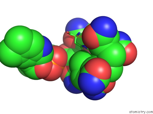
Mono view
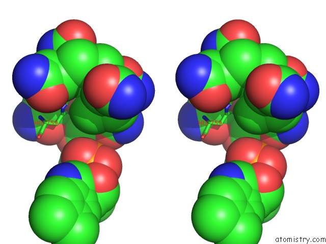
Stereo pair view
Mono view
Stereo pair view
A full contact list of Cobalt with other atoms in the Co binding
site number 1 of How A Protein Binds B12: A 3.O Angstrom X-Ray Structure of the B12-Binding Domains of Methionine Synthase within 5.0Å range:
|
Cobalt binding site 2 out of 2 in 1bmt
Go back to
Cobalt binding site 2 out
of 2 in the How A Protein Binds B12: A 3.O Angstrom X-Ray Structure of the B12-Binding Domains of Methionine Synthase
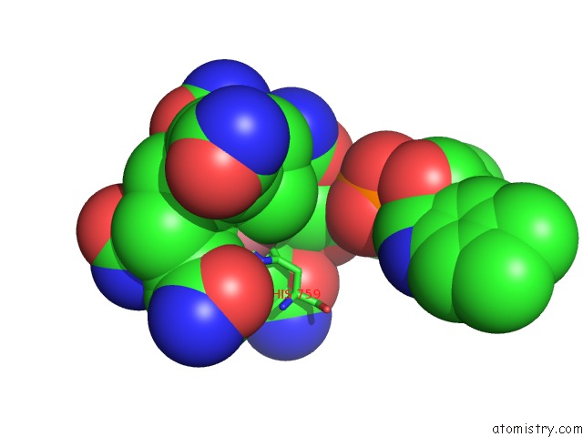
Mono view
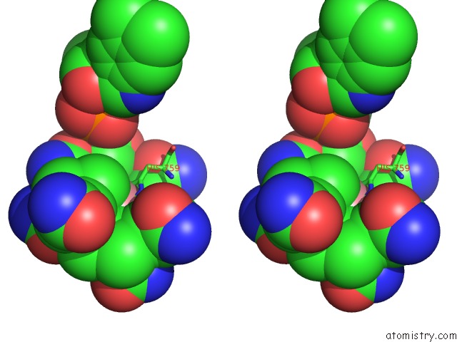
Stereo pair view
Mono view
Stereo pair view
A full contact list of Cobalt with other atoms in the Co binding
site number 2 of How A Protein Binds B12: A 3.O Angstrom X-Ray Structure of the B12-Binding Domains of Methionine Synthase within 5.0Å range:
|
Reference:
C.L.Drennan,
S.Huang,
J.T.Drummond,
R.G.Matthews,
M.L.Lidwig.
How A Protein Binds B12: A 3.0 A X-Ray Structure of B12-Binding Domains of Methionine Synthase. Science V. 266 1669 1994.
ISSN: ISSN 0036-8075
PubMed: 7992050
Page generated: Sun Jul 13 17:23:46 2025
ISSN: ISSN 0036-8075
PubMed: 7992050
Last articles
W in 7RCJW in 7XQW
W in 7VW6
W in 7T5A
W in 7OWZ
W in 7OV7
W in 7OVS
W in 7AX2
W in 7E5Z
W in 7A9M