Cobalt »
PDB 1a0c-1e1c »
1boa »
Cobalt in PDB 1boa: Human Methionine Aminopeptidase 2 Complexed with Angiogenesis Inhibitor Fumagillin
Enzymatic activity of Human Methionine Aminopeptidase 2 Complexed with Angiogenesis Inhibitor Fumagillin
All present enzymatic activity of Human Methionine Aminopeptidase 2 Complexed with Angiogenesis Inhibitor Fumagillin:
3.4.11.18;
3.4.11.18;
Protein crystallography data
The structure of Human Methionine Aminopeptidase 2 Complexed with Angiogenesis Inhibitor Fumagillin, PDB code: 1boa
was solved by
S.Liu,
J.Widom,
C.W.Kemp,
C.M.Crews,
J.C.Clardy,
with X-Ray Crystallography technique. A brief refinement statistics is given in the table below:
| Resolution Low / High (Å) | 25.00 / 1.80 |
| Space group | C 2 2 21 |
| Cell size a, b, c (Å), α, β, γ (°) | 90.700, 99.580, 101.950, 90.00, 90.00, 90.00 |
| R / Rfree (%) | 19.3 / 23.2 |
Cobalt Binding Sites:
The binding sites of Cobalt atom in the Human Methionine Aminopeptidase 2 Complexed with Angiogenesis Inhibitor Fumagillin
(pdb code 1boa). This binding sites where shown within
5.0 Angstroms radius around Cobalt atom.
In total 2 binding sites of Cobalt where determined in the Human Methionine Aminopeptidase 2 Complexed with Angiogenesis Inhibitor Fumagillin, PDB code: 1boa:
Jump to Cobalt binding site number: 1; 2;
In total 2 binding sites of Cobalt where determined in the Human Methionine Aminopeptidase 2 Complexed with Angiogenesis Inhibitor Fumagillin, PDB code: 1boa:
Jump to Cobalt binding site number: 1; 2;
Cobalt binding site 1 out of 2 in 1boa
Go back to
Cobalt binding site 1 out
of 2 in the Human Methionine Aminopeptidase 2 Complexed with Angiogenesis Inhibitor Fumagillin
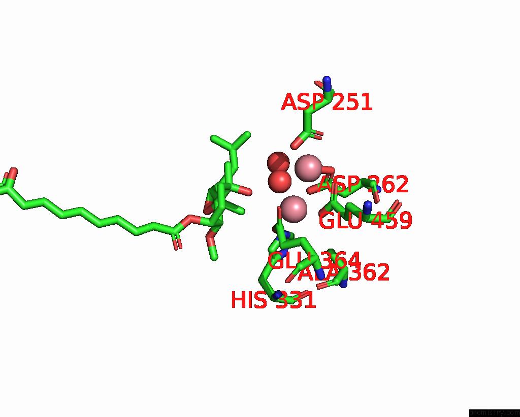
Mono view
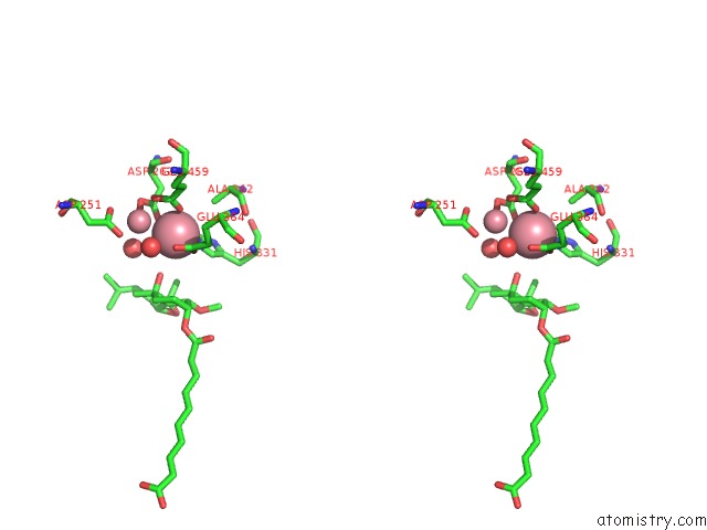
Stereo pair view
Mono view
Stereo pair view
A full contact list of Cobalt with other atoms in the Co binding
site number 1 of Human Methionine Aminopeptidase 2 Complexed with Angiogenesis Inhibitor Fumagillin within 5.0Å range:
|
Cobalt binding site 2 out of 2 in 1boa
Go back to
Cobalt binding site 2 out
of 2 in the Human Methionine Aminopeptidase 2 Complexed with Angiogenesis Inhibitor Fumagillin
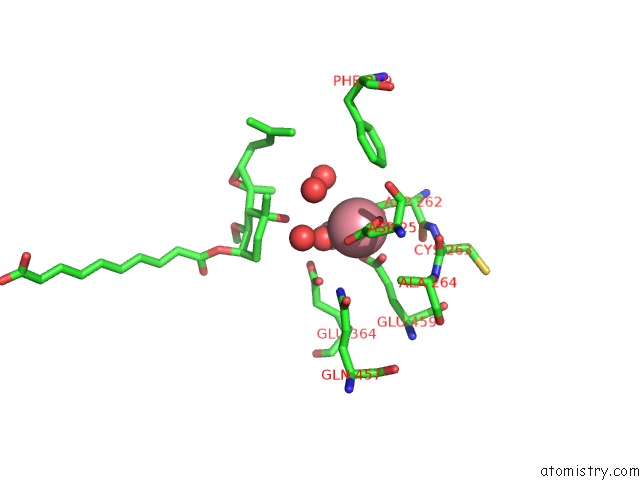
Mono view
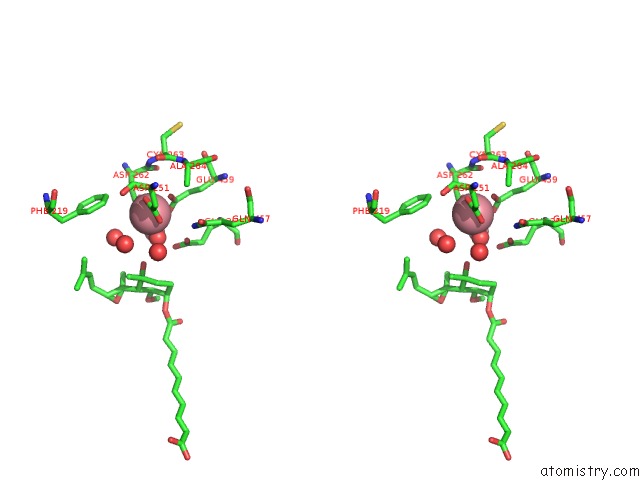
Stereo pair view
Mono view
Stereo pair view
A full contact list of Cobalt with other atoms in the Co binding
site number 2 of Human Methionine Aminopeptidase 2 Complexed with Angiogenesis Inhibitor Fumagillin within 5.0Å range:
|
Reference:
S.Liu,
J.Widom,
C.W.Kemp,
C.M.Crews,
J.Clardy.
Structure of Human Methionine Aminopeptidase-2 Complexed with Fumagillin. Science V. 282 1324 1998.
ISSN: ISSN 0036-8075
PubMed: 9812898
DOI: 10.1126/SCIENCE.282.5392.1324
Page generated: Sun Jul 13 17:24:02 2025
ISSN: ISSN 0036-8075
PubMed: 9812898
DOI: 10.1126/SCIENCE.282.5392.1324
Last articles
W in 7RCJW in 7XQW
W in 7VW6
W in 7T5A
W in 7OWZ
W in 7OV7
W in 7OVS
W in 7AX2
W in 7E5Z
W in 7A9M