Cobalt »
PDB 1e31-1hgw »
1gqj »
Cobalt in PDB 1gqj: Structure of Pseudomonas Cellulosa Alpha-D-Glucuronidase Complexed with Xylobiose
Enzymatic activity of Structure of Pseudomonas Cellulosa Alpha-D-Glucuronidase Complexed with Xylobiose
All present enzymatic activity of Structure of Pseudomonas Cellulosa Alpha-D-Glucuronidase Complexed with Xylobiose:
3.2.1.139;
3.2.1.139;
Protein crystallography data
The structure of Structure of Pseudomonas Cellulosa Alpha-D-Glucuronidase Complexed with Xylobiose, PDB code: 1gqj
was solved by
D.Nurizzo,
T.Nagy,
H.J.Gilbert,
G.J.Davies,
with X-Ray Crystallography technique. A brief refinement statistics is given in the table below:
| Resolution Low / High (Å) | 20.00 / 1.90 |
| Space group | P 1 |
| Cell size a, b, c (Å), α, β, γ (°) | 69.561, 74.609, 87.450, 115.23, 93.15, 109.17 |
| R / Rfree (%) | 17.2 / 22.1 |
Cobalt Binding Sites:
The binding sites of Cobalt atom in the Structure of Pseudomonas Cellulosa Alpha-D-Glucuronidase Complexed with Xylobiose
(pdb code 1gqj). This binding sites where shown within
5.0 Angstroms radius around Cobalt atom.
In total 8 binding sites of Cobalt where determined in the Structure of Pseudomonas Cellulosa Alpha-D-Glucuronidase Complexed with Xylobiose, PDB code: 1gqj:
Jump to Cobalt binding site number: 1; 2; 3; 4; 5; 6; 7; 8;
In total 8 binding sites of Cobalt where determined in the Structure of Pseudomonas Cellulosa Alpha-D-Glucuronidase Complexed with Xylobiose, PDB code: 1gqj:
Jump to Cobalt binding site number: 1; 2; 3; 4; 5; 6; 7; 8;
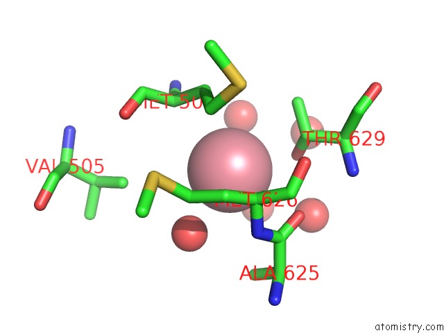
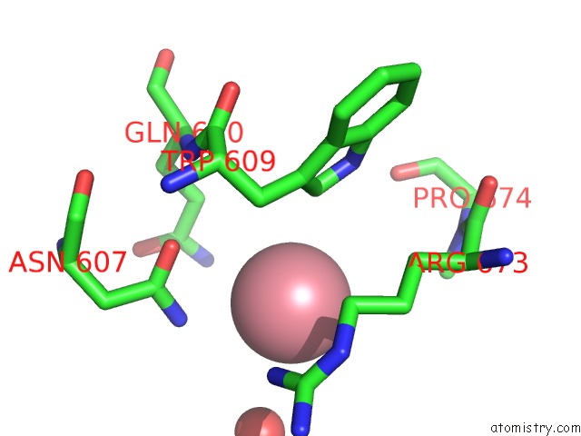
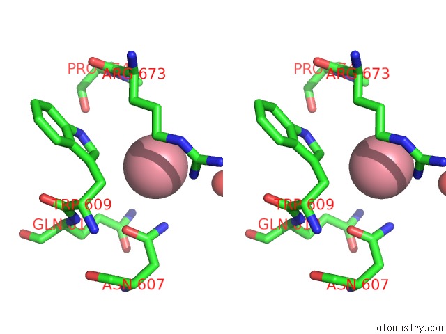
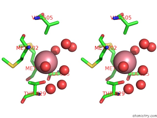
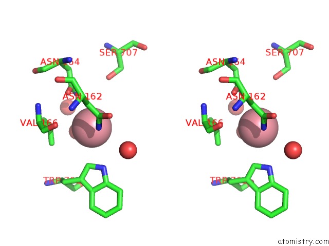
Cobalt binding site 1 out of 8 in 1gqj
Go back to
Cobalt binding site 1 out
of 8 in the Structure of Pseudomonas Cellulosa Alpha-D-Glucuronidase Complexed with Xylobiose
Mono view
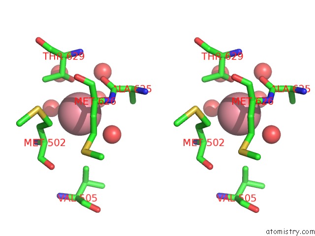
Stereo pair view
Mono view
Stereo pair view
A full contact list of Cobalt with other atoms in the Co binding
site number 1 of Structure of Pseudomonas Cellulosa Alpha-D-Glucuronidase Complexed with Xylobiose within 5.0Å range:
|
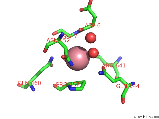
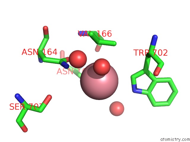
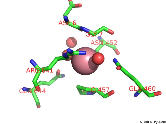
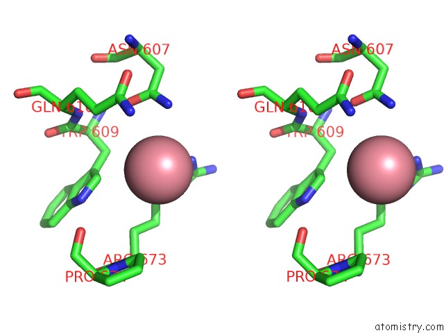
Cobalt binding site 2 out of 8 in 1gqj
Go back to
Cobalt binding site 2 out
of 8 in the Structure of Pseudomonas Cellulosa Alpha-D-Glucuronidase Complexed with Xylobiose
Mono view
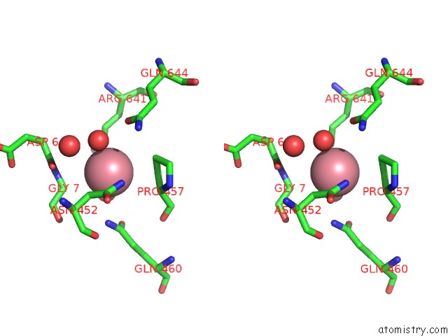
Stereo pair view
Mono view
Stereo pair view
A full contact list of Cobalt with other atoms in the Co binding
site number 2 of Structure of Pseudomonas Cellulosa Alpha-D-Glucuronidase Complexed with Xylobiose within 5.0Å range:
|
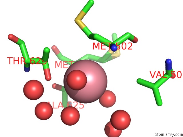
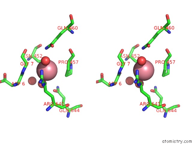
Cobalt binding site 3 out of 8 in 1gqj
Go back to
Cobalt binding site 3 out
of 8 in the Structure of Pseudomonas Cellulosa Alpha-D-Glucuronidase Complexed with Xylobiose
Mono view
Stereo pair view
Mono view
Stereo pair view
A full contact list of Cobalt with other atoms in the Co binding
site number 3 of Structure of Pseudomonas Cellulosa Alpha-D-Glucuronidase Complexed with Xylobiose within 5.0Å range:
|
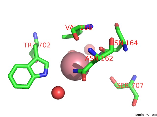
Cobalt binding site 4 out of 8 in 1gqj
Go back to
Cobalt binding site 4 out
of 8 in the Structure of Pseudomonas Cellulosa Alpha-D-Glucuronidase Complexed with Xylobiose
Mono view

Stereo pair view
Mono view
Stereo pair view
A full contact list of Cobalt with other atoms in the Co binding
site number 4 of Structure of Pseudomonas Cellulosa Alpha-D-Glucuronidase Complexed with Xylobiose within 5.0Å range:
|
Cobalt binding site 5 out of 8 in 1gqj
Go back to
Cobalt binding site 5 out
of 8 in the Structure of Pseudomonas Cellulosa Alpha-D-Glucuronidase Complexed with Xylobiose
Mono view
Stereo pair view
Mono view
Stereo pair view
A full contact list of Cobalt with other atoms in the Co binding
site number 5 of Structure of Pseudomonas Cellulosa Alpha-D-Glucuronidase Complexed with Xylobiose within 5.0Å range:
|
Cobalt binding site 6 out of 8 in 1gqj
Go back to
Cobalt binding site 6 out
of 8 in the Structure of Pseudomonas Cellulosa Alpha-D-Glucuronidase Complexed with Xylobiose
Mono view
Stereo pair view
Mono view
Stereo pair view
A full contact list of Cobalt with other atoms in the Co binding
site number 6 of Structure of Pseudomonas Cellulosa Alpha-D-Glucuronidase Complexed with Xylobiose within 5.0Å range:
|
Cobalt binding site 7 out of 8 in 1gqj
Go back to
Cobalt binding site 7 out
of 8 in the Structure of Pseudomonas Cellulosa Alpha-D-Glucuronidase Complexed with Xylobiose
Mono view
Stereo pair view
Mono view
Stereo pair view
A full contact list of Cobalt with other atoms in the Co binding
site number 7 of Structure of Pseudomonas Cellulosa Alpha-D-Glucuronidase Complexed with Xylobiose within 5.0Å range:
|
Cobalt binding site 8 out of 8 in 1gqj
Go back to
Cobalt binding site 8 out
of 8 in the Structure of Pseudomonas Cellulosa Alpha-D-Glucuronidase Complexed with Xylobiose

Mono view
Stereo pair view
Mono view
Stereo pair view
A full contact list of Cobalt with other atoms in the Co binding
site number 8 of Structure of Pseudomonas Cellulosa Alpha-D-Glucuronidase Complexed with Xylobiose within 5.0Å range:
|
Reference:
D.Nurizzo,
T.Nagy,
H.J.Gilbert,
G.J.Davies.
The Structural Basis For Catalysis and Specificity of the Pseudomonas Cellulosa Alpha-Glucuronidase, GLCA67A Structure V. 10 547 2002.
ISSN: ISSN 0969-2126
PubMed: 11937059
DOI: 10.1016/S0969-2126(02)00742-6
Page generated: Sun Jul 13 17:32:02 2025
ISSN: ISSN 0969-2126
PubMed: 11937059
DOI: 10.1016/S0969-2126(02)00742-6
Last articles
Mg in 4QZ4Mg in 4QZ3
Mg in 4QZ2
Mg in 4QZ0
Mg in 4QZ1
Mg in 4QYI
Mg in 4QYT
Mg in 4QYM
Mg in 4QY6
Mg in 4QXJ