Cobalt »
PDB 1hv9-1nr5 »
1mmf »
Cobalt in PDB 1mmf: Crystal Structure of Substrate Free Form of Glycerol Dehydratase
Enzymatic activity of Crystal Structure of Substrate Free Form of Glycerol Dehydratase
All present enzymatic activity of Crystal Structure of Substrate Free Form of Glycerol Dehydratase:
4.2.1.30;
4.2.1.30;
Protein crystallography data
The structure of Crystal Structure of Substrate Free Form of Glycerol Dehydratase, PDB code: 1mmf
was solved by
D.I.Liao,
G.Dotson,
I.Turner,
L.Reiss,
M.Emptage,
with X-Ray Crystallography technique. A brief refinement statistics is given in the table below:
| Resolution Low / High (Å) | 30.00 / 2.50 |
| Space group | P 1 21 1 |
| Cell size a, b, c (Å), α, β, γ (°) | 93.700, 110.070, 114.690, 90.00, 107.64, 90.00 |
| R / Rfree (%) | 22.8 / 26.9 |
Other elements in 1mmf:
The structure of Crystal Structure of Substrate Free Form of Glycerol Dehydratase also contains other interesting chemical elements:
| Potassium | (K) | 2 atoms |
Cobalt Binding Sites:
The binding sites of Cobalt atom in the Crystal Structure of Substrate Free Form of Glycerol Dehydratase
(pdb code 1mmf). This binding sites where shown within
5.0 Angstroms radius around Cobalt atom.
In total 2 binding sites of Cobalt where determined in the Crystal Structure of Substrate Free Form of Glycerol Dehydratase, PDB code: 1mmf:
Jump to Cobalt binding site number: 1; 2;
In total 2 binding sites of Cobalt where determined in the Crystal Structure of Substrate Free Form of Glycerol Dehydratase, PDB code: 1mmf:
Jump to Cobalt binding site number: 1; 2;
Cobalt binding site 1 out of 2 in 1mmf
Go back to
Cobalt binding site 1 out
of 2 in the Crystal Structure of Substrate Free Form of Glycerol Dehydratase
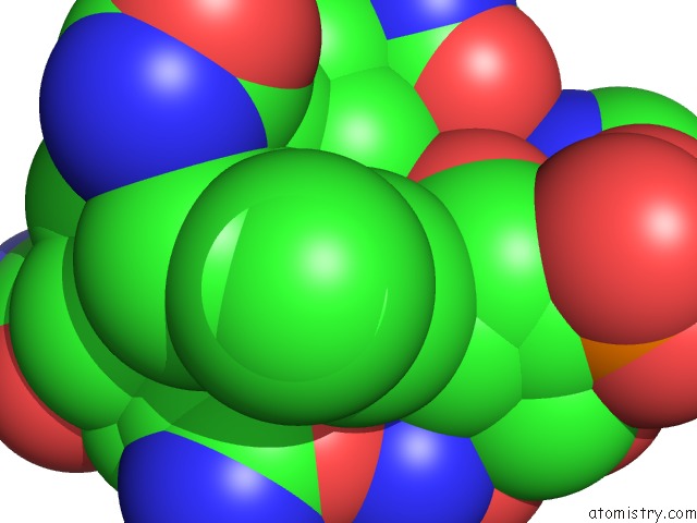
Mono view
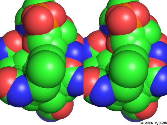
Stereo pair view
Mono view
Stereo pair view
A full contact list of Cobalt with other atoms in the Co binding
site number 1 of Crystal Structure of Substrate Free Form of Glycerol Dehydratase within 5.0Å range:
|
Cobalt binding site 2 out of 2 in 1mmf
Go back to
Cobalt binding site 2 out
of 2 in the Crystal Structure of Substrate Free Form of Glycerol Dehydratase
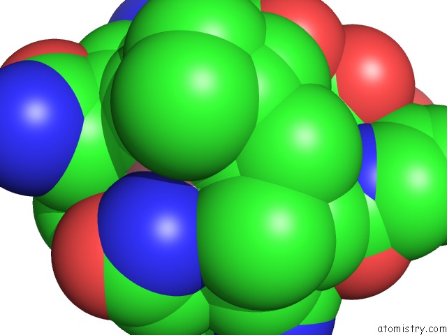
Mono view
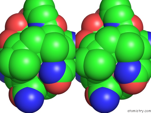
Stereo pair view
Mono view
Stereo pair view
A full contact list of Cobalt with other atoms in the Co binding
site number 2 of Crystal Structure of Substrate Free Form of Glycerol Dehydratase within 5.0Å range:
|
Reference:
D.I.Liao,
G.Dotson,
I.Turner,
L.Reiss,
M.Emptage.
Crystal Structure of Substrate Free Form of Glycerol Dehydratase J.Inorg.Biochem. V. 93 84 2003.
ISSN: ISSN 0162-0134
PubMed: 12538056
DOI: 10.1016/S0162-0134(02)00523-8
Page generated: Sun Jul 13 17:39:34 2025
ISSN: ISSN 0162-0134
PubMed: 12538056
DOI: 10.1016/S0162-0134(02)00523-8
Last articles
Mg in 5LMQMg in 5LMS
Mg in 5LMR
Mg in 5LMN
Mg in 5LMM
Mg in 5LLB
Mg in 5LMK
Mg in 5LMG
Mg in 5LLX
Mg in 5LM9