Cobalt »
PDB 1rrk-1vlx »
1sth »
Cobalt in PDB 1sth: Two Distinctly Different Metal Binding Modes Are Seen in X- Ray Crystal Structures of Staphylococcal Nuclease- Cobalt(II)-Nucleotide Complexes
Enzymatic activity of Two Distinctly Different Metal Binding Modes Are Seen in X- Ray Crystal Structures of Staphylococcal Nuclease- Cobalt(II)-Nucleotide Complexes
All present enzymatic activity of Two Distinctly Different Metal Binding Modes Are Seen in X- Ray Crystal Structures of Staphylococcal Nuclease- Cobalt(II)-Nucleotide Complexes:
3.1.31.1;
3.1.31.1;
Protein crystallography data
The structure of Two Distinctly Different Metal Binding Modes Are Seen in X- Ray Crystal Structures of Staphylococcal Nuclease- Cobalt(II)-Nucleotide Complexes, PDB code: 1sth
was solved by
P.J.Loll,
S.Quirk,
E.E.Lattman,
with X-Ray Crystallography technique. A brief refinement statistics is given in the table below:
| Resolution Low / High (Å) | 6.00 / 1.85 |
| Space group | P 41 |
| Cell size a, b, c (Å), α, β, γ (°) | 47.480, 47.480, 63.210, 90.00, 90.00, 90.00 |
| R / Rfree (%) | 17.4 / n/a |
Cobalt Binding Sites:
The binding sites of Cobalt atom in the Two Distinctly Different Metal Binding Modes Are Seen in X- Ray Crystal Structures of Staphylococcal Nuclease- Cobalt(II)-Nucleotide Complexes
(pdb code 1sth). This binding sites where shown within
5.0 Angstroms radius around Cobalt atom.
In total only one binding site of Cobalt was determined in the Two Distinctly Different Metal Binding Modes Are Seen in X- Ray Crystal Structures of Staphylococcal Nuclease- Cobalt(II)-Nucleotide Complexes, PDB code: 1sth:
In total only one binding site of Cobalt was determined in the Two Distinctly Different Metal Binding Modes Are Seen in X- Ray Crystal Structures of Staphylococcal Nuclease- Cobalt(II)-Nucleotide Complexes, PDB code: 1sth:
Cobalt binding site 1 out of 1 in 1sth
Go back to
Cobalt binding site 1 out
of 1 in the Two Distinctly Different Metal Binding Modes Are Seen in X- Ray Crystal Structures of Staphylococcal Nuclease- Cobalt(II)-Nucleotide Complexes
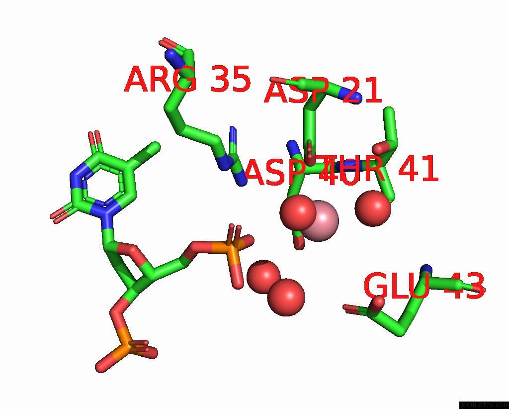
Mono view
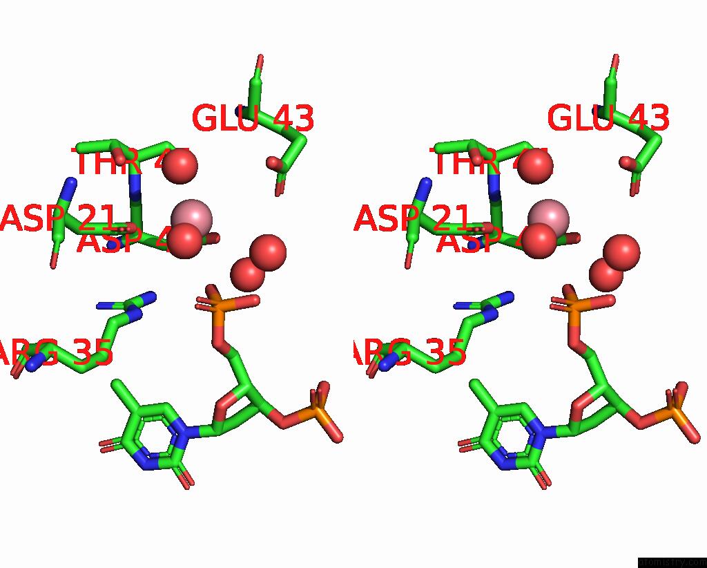
Stereo pair view
Mono view
Stereo pair view
A full contact list of Cobalt with other atoms in the Co binding
site number 1 of Two Distinctly Different Metal Binding Modes Are Seen in X- Ray Crystal Structures of Staphylococcal Nuclease- Cobalt(II)-Nucleotide Complexes within 5.0Å range:
|
Reference:
P.J.Loll,
S.Quirk,
E.E.Lattman,
R.M.Garavito.
X-Ray Crystal Structures of Staphylococcal Nuclease Complexed with the Competitive Inhibitor Cobalt(II) and Nucleotide. Biochemistry V. 34 4316 1995.
ISSN: ISSN 0006-2960
PubMed: 7703245
DOI: 10.1021/BI00013A021
Page generated: Sun Jul 13 17:48:03 2025
ISSN: ISSN 0006-2960
PubMed: 7703245
DOI: 10.1021/BI00013A021
Last articles
K in 4R33K in 4R2C
K in 4QXG
K in 4QRH
K in 4QNE
K in 4QGC
K in 4QKA
K in 4QE9
K in 4QG8
K in 4QK8