Cobalt »
PDB 1vz0-1zft »
1yog »
Cobalt in PDB 1yog: Cobalt Myoglobin (Deoxy)
Protein crystallography data
The structure of Cobalt Myoglobin (Deoxy), PDB code: 1yog
was solved by
E.A.Brucker,
G.N.Phillips Jr.,
with X-Ray Crystallography technique. A brief refinement statistics is given in the table below:
| Resolution Low / High (Å) | 5.00 / 1.65 |
| Space group | P 1 21 1 |
| Cell size a, b, c (Å), α, β, γ (°) | 64.710, 30.780, 34.870, 90.00, 106.08, 90.00 |
| R / Rfree (%) | 18.1 / n/a |
Cobalt Binding Sites:
The binding sites of Cobalt atom in the Cobalt Myoglobin (Deoxy)
(pdb code 1yog). This binding sites where shown within
5.0 Angstroms radius around Cobalt atom.
In total only one binding site of Cobalt was determined in the Cobalt Myoglobin (Deoxy), PDB code: 1yog:
In total only one binding site of Cobalt was determined in the Cobalt Myoglobin (Deoxy), PDB code: 1yog:
Cobalt binding site 1 out of 1 in 1yog
Go back to
Cobalt binding site 1 out
of 1 in the Cobalt Myoglobin (Deoxy)
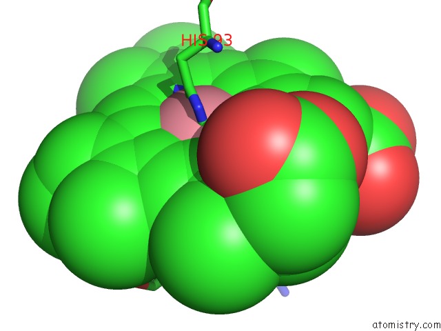
Mono view
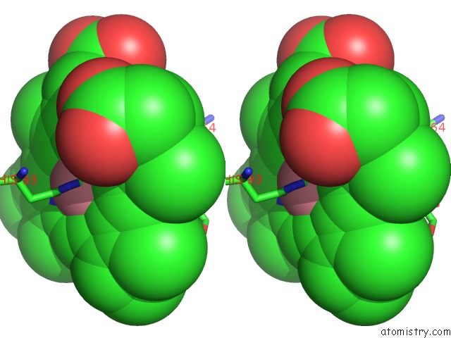
Stereo pair view
Mono view
Stereo pair view
A full contact list of Cobalt with other atoms in the Co binding
site number 1 of Cobalt Myoglobin (Deoxy) within 5.0Å range:
|
Reference:
E.A.Brucker,
J.S.Olson,
G.N.Phillips Jr.,
Y.Dou,
M.Ikeda-Saito.
High Resolution Crystal Structures of the Deoxy, Oxy, and Aquomet Forms of Cobalt Myoglobin. J.Biol.Chem. V. 271 25419 1996.
ISSN: ISSN 0021-9258
PubMed: 8810310
DOI: 10.1074/JBC.271.41.25419
Page generated: Sun Jul 13 17:59:52 2025
ISSN: ISSN 0021-9258
PubMed: 8810310
DOI: 10.1074/JBC.271.41.25419
Last articles
Na in 1VI6Na in 1VKG
Na in 1VMJ
Na in 1VMH
Na in 1VMF
Na in 1VLM
Na in 1VK1
Na in 1VIZ
Na in 1VEL
Na in 1VE8