Cobalt »
PDB 1zfv-2dfi »
2b3h »
Cobalt in PDB 2b3h: Crystal Structure of Human Methionine Aminopeptidase Type I with A Third Cobalt in the Active Site
Enzymatic activity of Crystal Structure of Human Methionine Aminopeptidase Type I with A Third Cobalt in the Active Site
All present enzymatic activity of Crystal Structure of Human Methionine Aminopeptidase Type I with A Third Cobalt in the Active Site:
3.4.11.18;
3.4.11.18;
Protein crystallography data
The structure of Crystal Structure of Human Methionine Aminopeptidase Type I with A Third Cobalt in the Active Site, PDB code: 2b3h
was solved by
A.Addlagatta,
X.Hu,
J.O.Liu,
B.W.Matthews,
with X-Ray Crystallography technique. A brief refinement statistics is given in the table below:
| Resolution Low / High (Å) | 20.00 / 1.10 |
| Space group | P 1 21 1 |
| Cell size a, b, c (Å), α, β, γ (°) | 47.290, 77.300, 48.340, 90.00, 91.03, 90.00 |
| R / Rfree (%) | 9.8 / 13.1 |
Other elements in 2b3h:
The structure of Crystal Structure of Human Methionine Aminopeptidase Type I with A Third Cobalt in the Active Site also contains other interesting chemical elements:
| Potassium | (K) | 1 atom |
| Chlorine | (Cl) | 1 atom |
Cobalt Binding Sites:
The binding sites of Cobalt atom in the Crystal Structure of Human Methionine Aminopeptidase Type I with A Third Cobalt in the Active Site
(pdb code 2b3h). This binding sites where shown within
5.0 Angstroms radius around Cobalt atom.
In total 4 binding sites of Cobalt where determined in the Crystal Structure of Human Methionine Aminopeptidase Type I with A Third Cobalt in the Active Site, PDB code: 2b3h:
Jump to Cobalt binding site number: 1; 2; 3; 4;
In total 4 binding sites of Cobalt where determined in the Crystal Structure of Human Methionine Aminopeptidase Type I with A Third Cobalt in the Active Site, PDB code: 2b3h:
Jump to Cobalt binding site number: 1; 2; 3; 4;
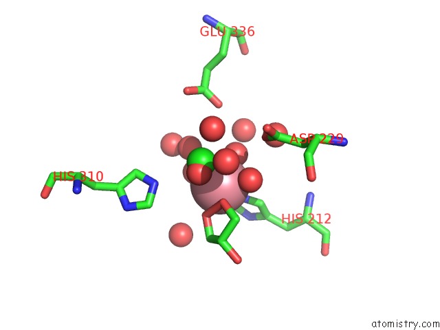
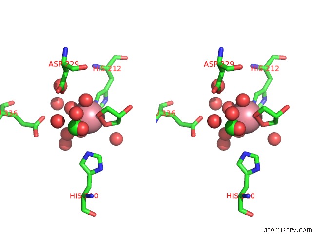
Cobalt binding site 1 out of 4 in 2b3h
Go back to
Cobalt binding site 1 out
of 4 in the Crystal Structure of Human Methionine Aminopeptidase Type I with A Third Cobalt in the Active Site
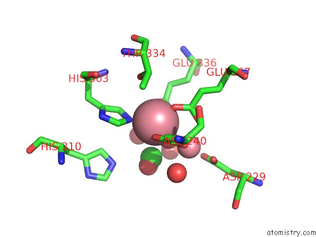
Mono view
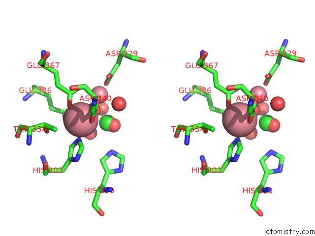
Stereo pair view
Mono view
Stereo pair view
A full contact list of Cobalt with other atoms in the Co binding
site number 1 of Crystal Structure of Human Methionine Aminopeptidase Type I with A Third Cobalt in the Active Site within 5.0Å range:
|
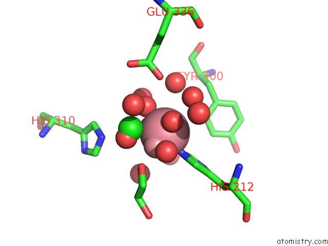
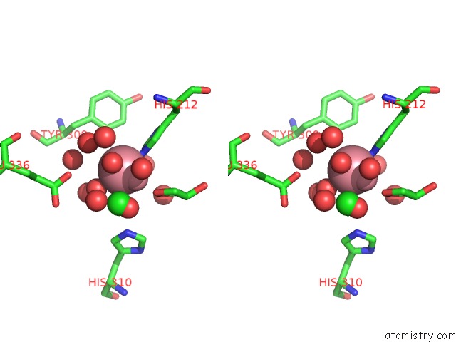
Cobalt binding site 2 out of 4 in 2b3h
Go back to
Cobalt binding site 2 out
of 4 in the Crystal Structure of Human Methionine Aminopeptidase Type I with A Third Cobalt in the Active Site
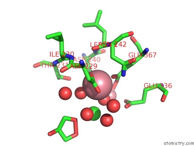
Mono view
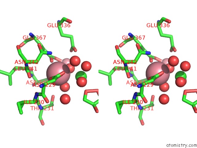
Stereo pair view
Mono view
Stereo pair view
A full contact list of Cobalt with other atoms in the Co binding
site number 2 of Crystal Structure of Human Methionine Aminopeptidase Type I with A Third Cobalt in the Active Site within 5.0Å range:
|
Cobalt binding site 3 out of 4 in 2b3h
Go back to
Cobalt binding site 3 out
of 4 in the Crystal Structure of Human Methionine Aminopeptidase Type I with A Third Cobalt in the Active Site
Mono view
Stereo pair view
Mono view
Stereo pair view
A full contact list of Cobalt with other atoms in the Co binding
site number 3 of Crystal Structure of Human Methionine Aminopeptidase Type I with A Third Cobalt in the Active Site within 5.0Å range:
|
Cobalt binding site 4 out of 4 in 2b3h
Go back to
Cobalt binding site 4 out
of 4 in the Crystal Structure of Human Methionine Aminopeptidase Type I with A Third Cobalt in the Active Site
Mono view
Stereo pair view
Mono view
Stereo pair view
A full contact list of Cobalt with other atoms in the Co binding
site number 4 of Crystal Structure of Human Methionine Aminopeptidase Type I with A Third Cobalt in the Active Site within 5.0Å range:
|
Reference:
A.Addlagatta,
X.Hu,
J.O.Liu,
B.W.Matthews.
Structural Basis For the Functional Differences Between Type I and Type II Human Methionine Aminopeptidases(,). Biochemistry V. 44 14741 2005.
ISSN: ISSN 0006-2960
PubMed: 16274222
DOI: 10.1021/BI051691K
Page generated: Sun Jul 13 18:02:14 2025
ISSN: ISSN 0006-2960
PubMed: 16274222
DOI: 10.1021/BI051691K
Last articles
Mg in 4DR5Mg in 4DUX
Mg in 4DUW
Mg in 4DUV
Mg in 4DUO
Mg in 4DUG
Mg in 4DTY
Mg in 4DTW
Mg in 4DTH
Mg in 4DTF