Cobalt »
PDB 2djl-2g6p »
2ero »
Cobalt in PDB 2ero: Crystal Structure of Vascular Apoptosis-Inducing Protein- 1(Orthorhombic Crystal Form)
Protein crystallography data
The structure of Crystal Structure of Vascular Apoptosis-Inducing Protein- 1(Orthorhombic Crystal Form), PDB code: 2ero
was solved by
S.Takeda,
T.Igarashi,
S.Araki,
with X-Ray Crystallography technique. A brief refinement statistics is given in the table below:
| Resolution Low / High (Å) | 50.00 / 2.50 |
| Space group | P 21 21 21 |
| Cell size a, b, c (Å), α, β, γ (°) | 86.710, 93.270, 137.740, 90.00, 90.00, 90.00 |
| R / Rfree (%) | 21.2 / 25.8 |
Other elements in 2ero:
The structure of Crystal Structure of Vascular Apoptosis-Inducing Protein- 1(Orthorhombic Crystal Form) also contains other interesting chemical elements:
| Calcium | (Ca) | 4 atoms |
| Zinc | (Zn) | 2 atoms |
Cobalt Binding Sites:
The binding sites of Cobalt atom in the Crystal Structure of Vascular Apoptosis-Inducing Protein- 1(Orthorhombic Crystal Form)
(pdb code 2ero). This binding sites where shown within
5.0 Angstroms radius around Cobalt atom.
In total only one binding site of Cobalt was determined in the Crystal Structure of Vascular Apoptosis-Inducing Protein- 1(Orthorhombic Crystal Form), PDB code: 2ero:
In total only one binding site of Cobalt was determined in the Crystal Structure of Vascular Apoptosis-Inducing Protein- 1(Orthorhombic Crystal Form), PDB code: 2ero:
Cobalt binding site 1 out of 1 in 2ero
Go back to
Cobalt binding site 1 out
of 1 in the Crystal Structure of Vascular Apoptosis-Inducing Protein- 1(Orthorhombic Crystal Form)
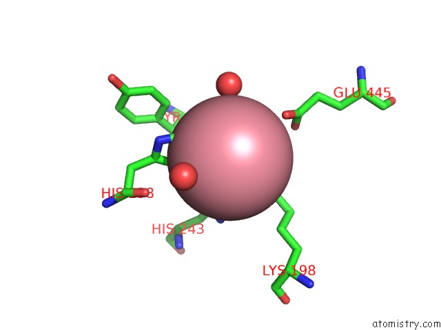
Mono view
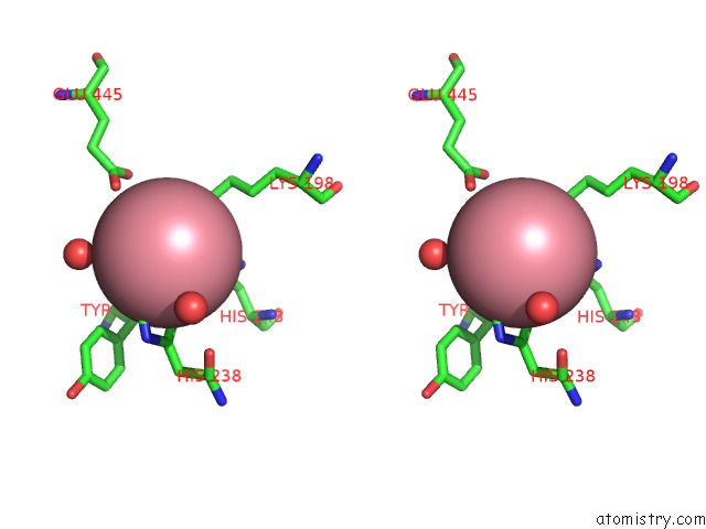
Stereo pair view
Mono view
Stereo pair view
A full contact list of Cobalt with other atoms in the Co binding
site number 1 of Crystal Structure of Vascular Apoptosis-Inducing Protein- 1(Orthorhombic Crystal Form) within 5.0Å range:
|
Reference:
S.Takeda,
T.Igarashi,
H.Mori,
S.Araki.
Crystal Structures of VAP1 Reveal Adams' Mdc Domain Architecture and Its Unique C-Shaped Scaffold Embo J. V. 25 2388 2006.
ISSN: ISSN 0261-4189
PubMed: 16688218
DOI: 10.1038/SJ.EMBOJ.7601131
Page generated: Sun Jul 13 18:12:35 2025
ISSN: ISSN 0261-4189
PubMed: 16688218
DOI: 10.1038/SJ.EMBOJ.7601131
Last articles
Fe in 8AIOFe in 8AMQ
Fe in 8AMP
Fe in 8AMO
Fe in 8AM5
Fe in 8ALJ
Fe in 8ALI
Fe in 8AJZ
Fe in 8AI6
Fe in 8AI5