Cobalt »
PDB 2g9c-2nq7 »
2hp5 »
Cobalt in PDB 2hp5: Crystal Structure of the Oxa-10 W154G Mutant at pH 7.0
Enzymatic activity of Crystal Structure of the Oxa-10 W154G Mutant at pH 7.0
All present enzymatic activity of Crystal Structure of the Oxa-10 W154G Mutant at pH 7.0:
3.5.2.6;
3.5.2.6;
Protein crystallography data
The structure of Crystal Structure of the Oxa-10 W154G Mutant at pH 7.0, PDB code: 2hp5
was solved by
F.Kerff,
C.Falzone,
R.Herman,
E.Sauvage,
P.Charlier,
with X-Ray Crystallography technique. A brief refinement statistics is given in the table below:
| Resolution Low / High (Å) | 46.47 / 2.70 |
| Space group | P 1 21 1 |
| Cell size a, b, c (Å), α, β, γ (°) | 47.110, 125.400, 92.360, 90.00, 99.80, 90.00 |
| R / Rfree (%) | 21 / 26.7 |
Cobalt Binding Sites:
The binding sites of Cobalt atom in the Crystal Structure of the Oxa-10 W154G Mutant at pH 7.0
(pdb code 2hp5). This binding sites where shown within
5.0 Angstroms radius around Cobalt atom.
In total 4 binding sites of Cobalt where determined in the Crystal Structure of the Oxa-10 W154G Mutant at pH 7.0, PDB code: 2hp5:
Jump to Cobalt binding site number: 1; 2; 3; 4;
In total 4 binding sites of Cobalt where determined in the Crystal Structure of the Oxa-10 W154G Mutant at pH 7.0, PDB code: 2hp5:
Jump to Cobalt binding site number: 1; 2; 3; 4;
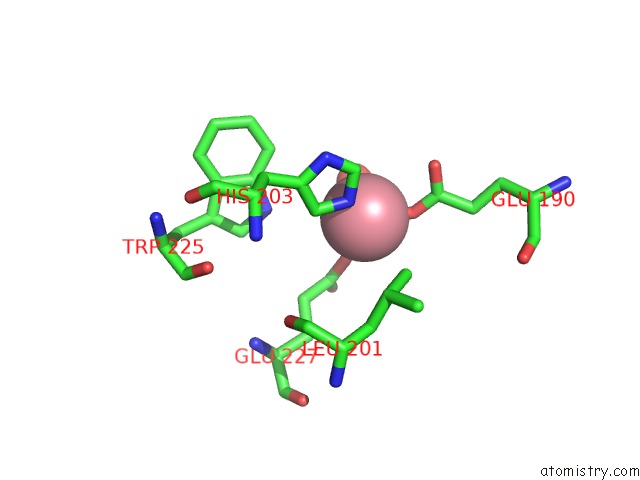
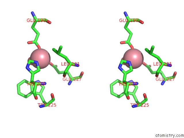
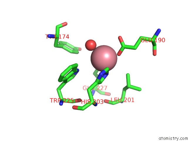
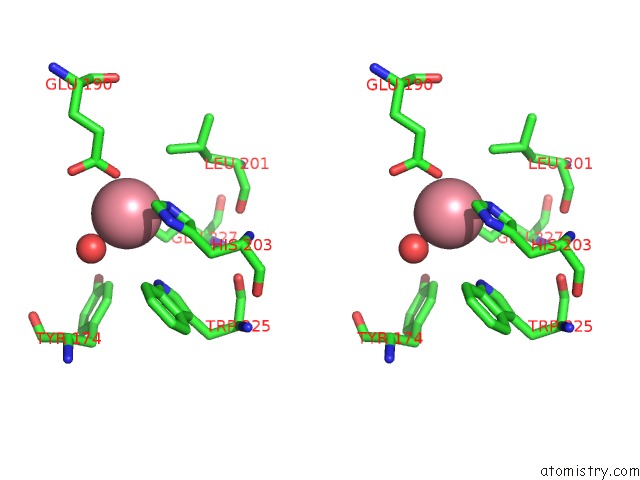
Cobalt binding site 1 out of 4 in 2hp5
Go back to
Cobalt binding site 1 out
of 4 in the Crystal Structure of the Oxa-10 W154G Mutant at pH 7.0
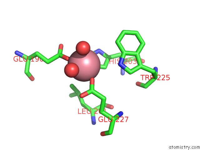
Mono view
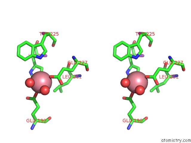
Stereo pair view
Mono view
Stereo pair view
A full contact list of Cobalt with other atoms in the Co binding
site number 1 of Crystal Structure of the Oxa-10 W154G Mutant at pH 7.0 within 5.0Å range:
|
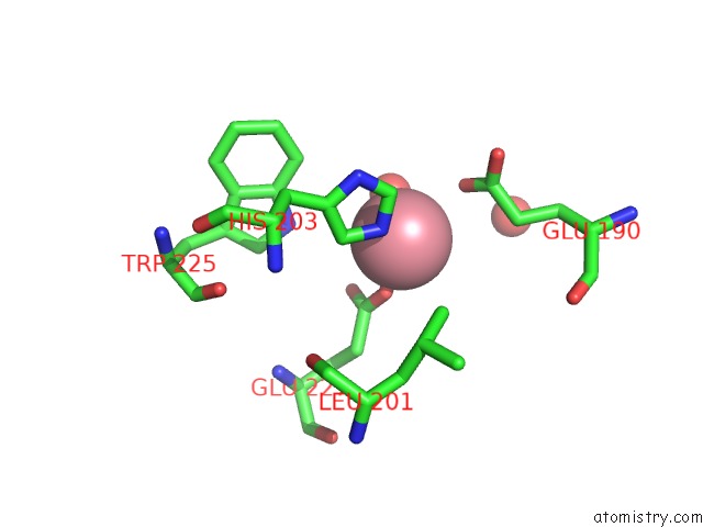
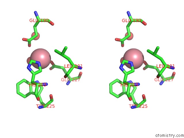
Cobalt binding site 2 out of 4 in 2hp5
Go back to
Cobalt binding site 2 out
of 4 in the Crystal Structure of the Oxa-10 W154G Mutant at pH 7.0
Mono view
Stereo pair view
Mono view
Stereo pair view
A full contact list of Cobalt with other atoms in the Co binding
site number 2 of Crystal Structure of the Oxa-10 W154G Mutant at pH 7.0 within 5.0Å range:
|
Cobalt binding site 3 out of 4 in 2hp5
Go back to
Cobalt binding site 3 out
of 4 in the Crystal Structure of the Oxa-10 W154G Mutant at pH 7.0
Mono view
Stereo pair view
Mono view
Stereo pair view
A full contact list of Cobalt with other atoms in the Co binding
site number 3 of Crystal Structure of the Oxa-10 W154G Mutant at pH 7.0 within 5.0Å range:
|
Cobalt binding site 4 out of 4 in 2hp5
Go back to
Cobalt binding site 4 out
of 4 in the Crystal Structure of the Oxa-10 W154G Mutant at pH 7.0
Mono view
Stereo pair view
Mono view
Stereo pair view
A full contact list of Cobalt with other atoms in the Co binding
site number 4 of Crystal Structure of the Oxa-10 W154G Mutant at pH 7.0 within 5.0Å range:
|
Reference:
S.Baurin,
L.Vercheval,
F.Bouillenne,
C.Falzone,
A.Brans,
L.Jacquamet,
J.L.Ferrer,
E.Sauvage,
D.Dehareng,
J.M.Frere,
P.Charlier,
M.Galleni,
F.Kerff.
Critical Role of Tryptophan 154 For the Activity and Stability of Class D Beta-Lactamases. Biochemistry V. 48 11252 2009.
ISSN: ISSN 0006-2960
PubMed: 19860471
DOI: 10.1021/BI901548C
Page generated: Sun Jul 13 18:18:11 2025
ISSN: ISSN 0006-2960
PubMed: 19860471
DOI: 10.1021/BI901548C
Last articles
Mg in 6JLJMg in 6JIL
Mg in 6JKM
Mg in 6JJW
Mg in 6JJU
Mg in 6JJ9
Mg in 6JD2
Mg in 6JJ8
Mg in 6JIM
Mg in 6JD1