Cobalt »
PDB 2g9c-2nq7 »
2icv »
Cobalt in PDB 2icv: Kinetic and Crystallographic Studies of A Redesigned Manganese-Binding Site in Cytochrome C Peroxidase
Enzymatic activity of Kinetic and Crystallographic Studies of A Redesigned Manganese-Binding Site in Cytochrome C Peroxidase
All present enzymatic activity of Kinetic and Crystallographic Studies of A Redesigned Manganese-Binding Site in Cytochrome C Peroxidase:
1.11.1.5;
1.11.1.5;
Protein crystallography data
The structure of Kinetic and Crystallographic Studies of A Redesigned Manganese-Binding Site in Cytochrome C Peroxidase, PDB code: 2icv
was solved by
T.Pfister,
A.Y.Mirarefi,
A.J.Gengenbach,
X.Zhao,
C.D.N.Conaster,
Y.G.Gao,
H.Robinson,
C.F.Zukoski,
A.H.J.Wang,
Y.Lu,
with X-Ray Crystallography technique. A brief refinement statistics is given in the table below:
| Resolution Low / High (Å) | 10.00 / 1.60 |
| Space group | P 21 21 21 |
| Cell size a, b, c (Å), α, β, γ (°) | 43.958, 52.477, 136.250, 90.00, 90.00, 90.00 |
| R / Rfree (%) | 22.3 / 25.7 |
Other elements in 2icv:
The structure of Kinetic and Crystallographic Studies of A Redesigned Manganese-Binding Site in Cytochrome C Peroxidase also contains other interesting chemical elements:
| Iron | (Fe) | 1 atom |
Cobalt Binding Sites:
The binding sites of Cobalt atom in the Kinetic and Crystallographic Studies of A Redesigned Manganese-Binding Site in Cytochrome C Peroxidase
(pdb code 2icv). This binding sites where shown within
5.0 Angstroms radius around Cobalt atom.
In total only one binding site of Cobalt was determined in the Kinetic and Crystallographic Studies of A Redesigned Manganese-Binding Site in Cytochrome C Peroxidase, PDB code: 2icv:
In total only one binding site of Cobalt was determined in the Kinetic and Crystallographic Studies of A Redesigned Manganese-Binding Site in Cytochrome C Peroxidase, PDB code: 2icv:
Cobalt binding site 1 out of 1 in 2icv
Go back to
Cobalt binding site 1 out
of 1 in the Kinetic and Crystallographic Studies of A Redesigned Manganese-Binding Site in Cytochrome C Peroxidase
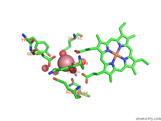
Mono view
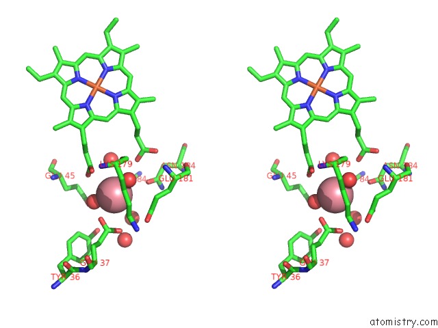
Stereo pair view
Mono view
Stereo pair view
A full contact list of Cobalt with other atoms in the Co binding
site number 1 of Kinetic and Crystallographic Studies of A Redesigned Manganese-Binding Site in Cytochrome C Peroxidase within 5.0Å range:
|
Reference:
T.D.Pfister,
A.Y.Mirarefi,
A.J.Gengenbach,
X.Zhao,
C.Danstrom,
N.Conatser,
Y.-G.Gao,
H.Robinson,
C.F.Zukoski,
A.H.-J.Wang,
Y.Lu.
Kinetic and Crystallographic Studies of A Redesigned Manganese-Binding Site in Cytochrome C Peroxidase J.Biol.Inorg.Chem. V. 12 126 2007.
ISSN: ISSN 0949-8257
PubMed: 17021923
DOI: 10.1007/S00775-006-0171-0
Page generated: Sun Jul 13 18:18:38 2025
ISSN: ISSN 0949-8257
PubMed: 17021923
DOI: 10.1007/S00775-006-0171-0
Last articles
K in 4AE4K in 4A69
K in 4A0M
K in 4A9K
K in 4A3U
K in 4A49
K in 4A1T
K in 3ZQA
K in 4A1V
K in 4A1O