Cobalt »
PDB 2r5v-2xwp »
2xts »
Cobalt in PDB 2xts: Crystal Structure of the Sulfane Dehydrogenase Soxcd From Paracoccus Pantotrophus
Enzymatic activity of Crystal Structure of the Sulfane Dehydrogenase Soxcd From Paracoccus Pantotrophus
All present enzymatic activity of Crystal Structure of the Sulfane Dehydrogenase Soxcd From Paracoccus Pantotrophus:
1.8.2.1;
1.8.2.1;
Protein crystallography data
The structure of Crystal Structure of the Sulfane Dehydrogenase Soxcd From Paracoccus Pantotrophus, PDB code: 2xts
was solved by
U.Zander,
A.Faust,
B.U.Klink,
D.De Sanctis,
S.Panjikar,
A.Quentmeier,
F.Bardischewski,
C.G.Friedrich,
A.J.Scheidig,
with X-Ray Crystallography technique. A brief refinement statistics is given in the table below:
| Resolution Low / High (Å) | 50.01 / 1.33 |
| Space group | P 31 |
| Cell size a, b, c (Å), α, β, γ (°) | 122.970, 122.970, 76.390, 90.00, 90.00, 120.00 |
| R / Rfree (%) | 10.699 / 11.11 |
Other elements in 2xts:
The structure of Crystal Structure of the Sulfane Dehydrogenase Soxcd From Paracoccus Pantotrophus also contains other interesting chemical elements:
| Molybdenum | (Mo) | 2 atoms |
| Iron | (Fe) | 2 atoms |
| Calcium | (Ca) | 6 atoms |
Cobalt Binding Sites:
The binding sites of Cobalt atom in the Crystal Structure of the Sulfane Dehydrogenase Soxcd From Paracoccus Pantotrophus
(pdb code 2xts). This binding sites where shown within
5.0 Angstroms radius around Cobalt atom.
In total 2 binding sites of Cobalt where determined in the Crystal Structure of the Sulfane Dehydrogenase Soxcd From Paracoccus Pantotrophus, PDB code: 2xts:
Jump to Cobalt binding site number: 1; 2;
In total 2 binding sites of Cobalt where determined in the Crystal Structure of the Sulfane Dehydrogenase Soxcd From Paracoccus Pantotrophus, PDB code: 2xts:
Jump to Cobalt binding site number: 1; 2;
Cobalt binding site 1 out of 2 in 2xts
Go back to
Cobalt binding site 1 out
of 2 in the Crystal Structure of the Sulfane Dehydrogenase Soxcd From Paracoccus Pantotrophus
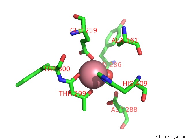
Mono view
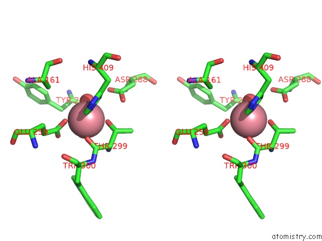
Stereo pair view
Mono view
Stereo pair view
A full contact list of Cobalt with other atoms in the Co binding
site number 1 of Crystal Structure of the Sulfane Dehydrogenase Soxcd From Paracoccus Pantotrophus within 5.0Å range:
|
Cobalt binding site 2 out of 2 in 2xts
Go back to
Cobalt binding site 2 out
of 2 in the Crystal Structure of the Sulfane Dehydrogenase Soxcd From Paracoccus Pantotrophus
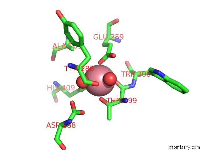
Mono view
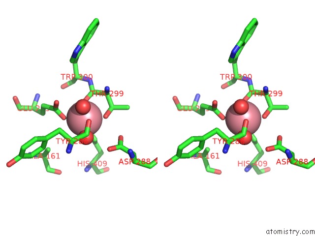
Stereo pair view
Mono view
Stereo pair view
A full contact list of Cobalt with other atoms in the Co binding
site number 2 of Crystal Structure of the Sulfane Dehydrogenase Soxcd From Paracoccus Pantotrophus within 5.0Å range:
|
Reference:
U.Zander,
A.Faust,
B.U.Klink,
D.De Sanctis,
S.Panjikar,
A.Quentmeier,
F.Bardischewsky,
C.G.Friedrich,
A.J.Scheidig.
Structural Basis For the Oxidation of Protein- Bound Sulfur By the Sulfur Cycle Molybdohemo- Enzyme Sulfane Dehydrogenase Soxcd. J.Biol.Chem. V. 286 8349 2011.
ISSN: ISSN 0021-9258
PubMed: 21147779
DOI: 10.1074/JBC.M110.193631
Page generated: Sun Jul 13 18:37:30 2025
ISSN: ISSN 0021-9258
PubMed: 21147779
DOI: 10.1074/JBC.M110.193631
Last articles
Mg in 8ZWQMg in 8ZUT
Mg in 8ZVC
Mg in 8ZUS
Mg in 8ZTZ
Mg in 8ZUQ
Mg in 8ZK2
Mg in 8ZUP
Mg in 8ZTF
Mg in 8ZTA