Cobalt »
PDB 2xwq-3bbi »
3abr »
Cobalt in PDB 3abr: Crystal Structure of Ethanolamine Ammonia-Lyase From Escherichia Coli Complexed with Cn-Cbl (Substrate-Free Form)
Enzymatic activity of Crystal Structure of Ethanolamine Ammonia-Lyase From Escherichia Coli Complexed with Cn-Cbl (Substrate-Free Form)
All present enzymatic activity of Crystal Structure of Ethanolamine Ammonia-Lyase From Escherichia Coli Complexed with Cn-Cbl (Substrate-Free Form):
4.3.1.7;
4.3.1.7;
Protein crystallography data
The structure of Crystal Structure of Ethanolamine Ammonia-Lyase From Escherichia Coli Complexed with Cn-Cbl (Substrate-Free Form), PDB code: 3abr
was solved by
N.Shibata,
with X-Ray Crystallography technique. A brief refinement statistics is given in the table below:
| Resolution Low / High (Å) | 42.29 / 2.10 |
| Space group | P 63 |
| Cell size a, b, c (Å), α, β, γ (°) | 244.120, 244.120, 77.150, 90.00, 90.00, 120.00 |
| R / Rfree (%) | 24.8 / 28.5 |
Other elements in 3abr:
The structure of Crystal Structure of Ethanolamine Ammonia-Lyase From Escherichia Coli Complexed with Cn-Cbl (Substrate-Free Form) also contains other interesting chemical elements:
| Sodium | (Na) | 7 atoms |
Cobalt Binding Sites:
The binding sites of Cobalt atom in the Crystal Structure of Ethanolamine Ammonia-Lyase From Escherichia Coli Complexed with Cn-Cbl (Substrate-Free Form)
(pdb code 3abr). This binding sites where shown within
5.0 Angstroms radius around Cobalt atom.
In total 2 binding sites of Cobalt where determined in the Crystal Structure of Ethanolamine Ammonia-Lyase From Escherichia Coli Complexed with Cn-Cbl (Substrate-Free Form), PDB code: 3abr:
Jump to Cobalt binding site number: 1; 2;
In total 2 binding sites of Cobalt where determined in the Crystal Structure of Ethanolamine Ammonia-Lyase From Escherichia Coli Complexed with Cn-Cbl (Substrate-Free Form), PDB code: 3abr:
Jump to Cobalt binding site number: 1; 2;
Cobalt binding site 1 out of 2 in 3abr
Go back to
Cobalt binding site 1 out
of 2 in the Crystal Structure of Ethanolamine Ammonia-Lyase From Escherichia Coli Complexed with Cn-Cbl (Substrate-Free Form)
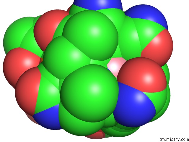
Mono view
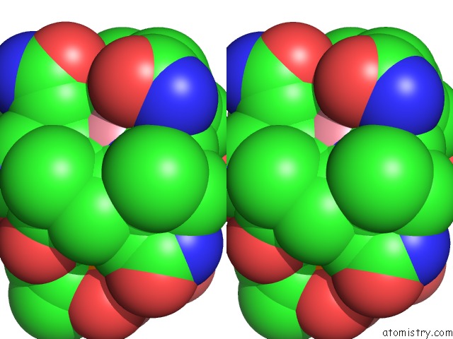
Stereo pair view
Mono view
Stereo pair view
A full contact list of Cobalt with other atoms in the Co binding
site number 1 of Crystal Structure of Ethanolamine Ammonia-Lyase From Escherichia Coli Complexed with Cn-Cbl (Substrate-Free Form) within 5.0Å range:
|
Cobalt binding site 2 out of 2 in 3abr
Go back to
Cobalt binding site 2 out
of 2 in the Crystal Structure of Ethanolamine Ammonia-Lyase From Escherichia Coli Complexed with Cn-Cbl (Substrate-Free Form)
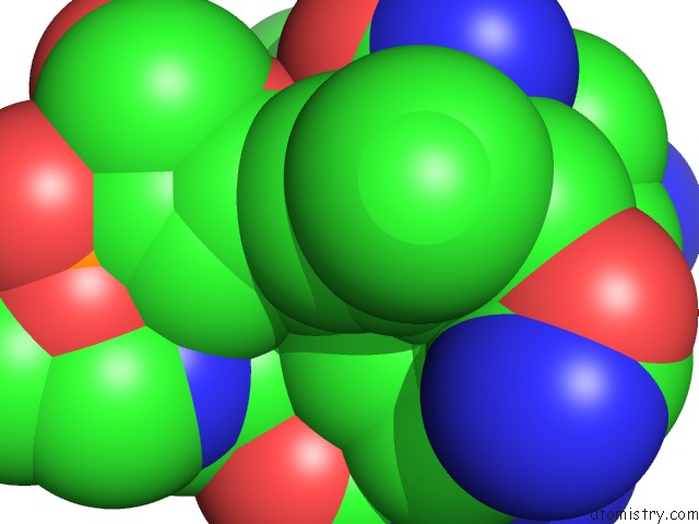
Mono view
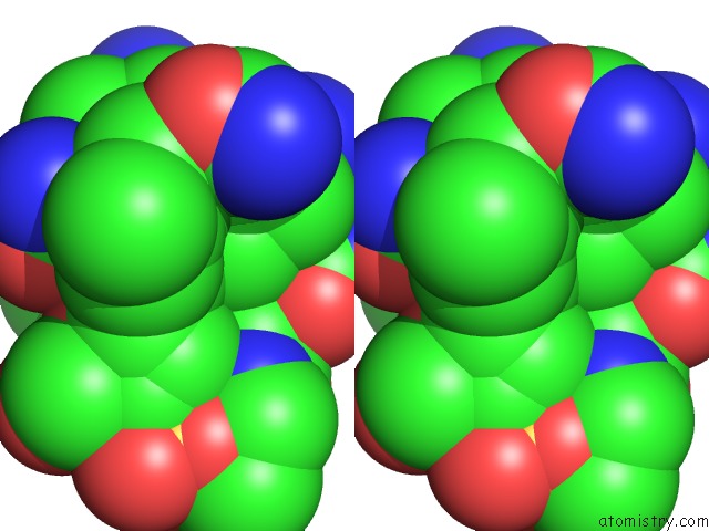
Stereo pair view
Mono view
Stereo pair view
A full contact list of Cobalt with other atoms in the Co binding
site number 2 of Crystal Structure of Ethanolamine Ammonia-Lyase From Escherichia Coli Complexed with Cn-Cbl (Substrate-Free Form) within 5.0Å range:
|
Reference:
N.Shibata,
H.Tamagaki,
N.Hieda,
K.Akita,
H.Komori,
Y.Shomura,
S.Terawaki,
K.Mori,
N.Yasuoka,
Y.Higuchi,
T.Toraya.
Crystal Structures of Ethanolamine Ammonia-Lyase Complexed with Coenzyme B12 Analogs and Substrates. J.Biol.Chem. V. 285 26484 2010.
ISSN: ISSN 0021-9258
PubMed: 20519496
DOI: 10.1074/JBC.M110.125112
Page generated: Sun Jul 13 18:42:36 2025
ISSN: ISSN 0021-9258
PubMed: 20519496
DOI: 10.1074/JBC.M110.125112
Last articles
Mg in 5KUTMg in 5KU1
Mg in 5KUE
Mg in 5KTY
Mg in 5KUJ
Mg in 5KTJ
Mg in 5KOZ
Mg in 5KT2
Mg in 5KTD
Mg in 5KT6