Cobalt »
PDB 3bbk-3ger »
3ezx »
Cobalt in PDB 3ezx: Structure of Methanosarcina Barkeri Monomethylamine Corrinoid Protein
Protein crystallography data
The structure of Structure of Methanosarcina Barkeri Monomethylamine Corrinoid Protein, PDB code: 3ezx
was solved by
R.Jain,
with X-Ray Crystallography technique. A brief refinement statistics is given in the table below:
| Resolution Low / High (Å) | 45.58 / 2.56 |
| Space group | C 2 2 21 |
| Cell size a, b, c (Å), α, β, γ (°) | 67.970, 77.710, 100.360, 90.00, 90.00, 90.00 |
| R / Rfree (%) | 20.5 / 26.2 |
Other elements in 3ezx:
The structure of Structure of Methanosarcina Barkeri Monomethylamine Corrinoid Protein also contains other interesting chemical elements:
| Magnesium | (Mg) | 1 atom |
Cobalt Binding Sites:
The binding sites of Cobalt atom in the Structure of Methanosarcina Barkeri Monomethylamine Corrinoid Protein
(pdb code 3ezx). This binding sites where shown within
5.0 Angstroms radius around Cobalt atom.
In total only one binding site of Cobalt was determined in the Structure of Methanosarcina Barkeri Monomethylamine Corrinoid Protein, PDB code: 3ezx:
In total only one binding site of Cobalt was determined in the Structure of Methanosarcina Barkeri Monomethylamine Corrinoid Protein, PDB code: 3ezx:
Cobalt binding site 1 out of 1 in 3ezx
Go back to
Cobalt binding site 1 out
of 1 in the Structure of Methanosarcina Barkeri Monomethylamine Corrinoid Protein
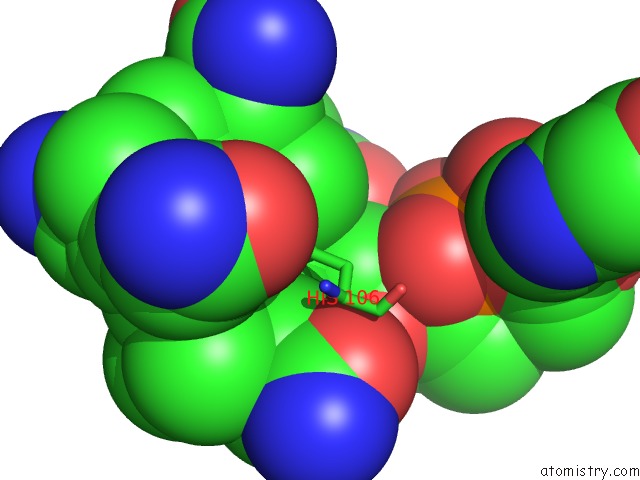
Mono view
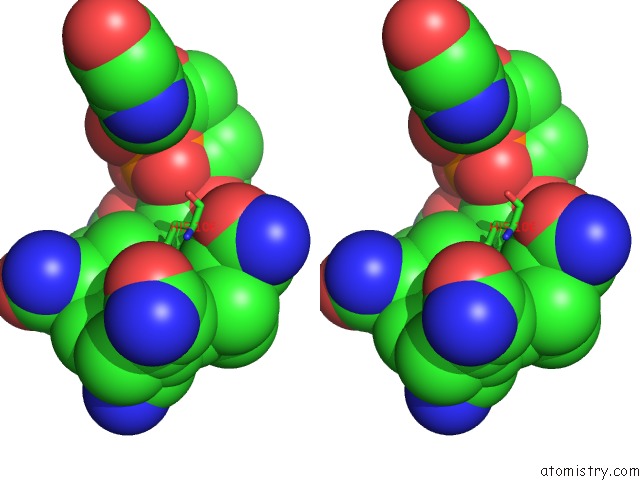
Stereo pair view
Mono view
Stereo pair view
A full contact list of Cobalt with other atoms in the Co binding
site number 1 of Structure of Methanosarcina Barkeri Monomethylamine Corrinoid Protein within 5.0Å range:
|
Reference:
R.Jain,
B.Hao,
J.A.Soares,
L.Zhang,
K.Green-Church,
X.Li,
J.A.Krzycki,
M.K.Chan.
Structure of Methanosarcina Barkeri Monomethylamine Corrinoid Protein To Be Published.
Page generated: Sun Jul 13 18:49:02 2025
Last articles
Mg in 1W55Mg in 1W54
Mg in 1W4B
Mg in 1W49
Mg in 1W46
Mg in 1W2Y
Mg in 1VQN
Mg in 1W25
Mg in 1W23
Mg in 1W1Z