Cobalt »
PDB 3igz-3mcr »
3isq »
Cobalt in PDB 3isq: Crystal Structure of Human 4-Hydroxyphenylpyruvate Dioxygenase
Enzymatic activity of Crystal Structure of Human 4-Hydroxyphenylpyruvate Dioxygenase
All present enzymatic activity of Crystal Structure of Human 4-Hydroxyphenylpyruvate Dioxygenase:
1.13.11.27;
1.13.11.27;
Protein crystallography data
The structure of Crystal Structure of Human 4-Hydroxyphenylpyruvate Dioxygenase, PDB code: 3isq
was solved by
E.S.Pilka,
N.Shafqat,
R.Cocking,
J.E.Bray,
T.Krojer,
A.C.W.Pike,
F.Vondelft,
W.W.Yue,
C.H.Arrowsmith,
J.Weigelt,
A.Edwards,
C.Bountra,
U.Oppermann,
K.L.Kavanagh,
Structural Genomics Consortium (Sgc),
with X-Ray Crystallography technique. A brief refinement statistics is given in the table below:
| Resolution Low / High (Å) | 49.60 / 1.75 |
| Space group | P 31 2 1 |
| Cell size a, b, c (Å), α, β, γ (°) | 99.206, 99.206, 87.726, 90.00, 90.00, 120.00 |
| R / Rfree (%) | 14.4 / 17.4 |
Other elements in 3isq:
The structure of Crystal Structure of Human 4-Hydroxyphenylpyruvate Dioxygenase also contains other interesting chemical elements:
| Chlorine | (Cl) | 1 atom |
| Sodium | (Na) | 1 atom |
Cobalt Binding Sites:
The binding sites of Cobalt atom in the Crystal Structure of Human 4-Hydroxyphenylpyruvate Dioxygenase
(pdb code 3isq). This binding sites where shown within
5.0 Angstroms radius around Cobalt atom.
In total only one binding site of Cobalt was determined in the Crystal Structure of Human 4-Hydroxyphenylpyruvate Dioxygenase, PDB code: 3isq:
In total only one binding site of Cobalt was determined in the Crystal Structure of Human 4-Hydroxyphenylpyruvate Dioxygenase, PDB code: 3isq:
Cobalt binding site 1 out of 1 in 3isq
Go back to
Cobalt binding site 1 out
of 1 in the Crystal Structure of Human 4-Hydroxyphenylpyruvate Dioxygenase
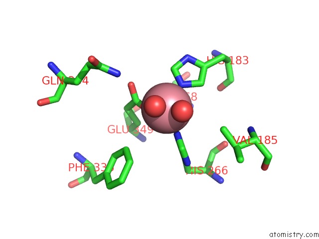
Mono view
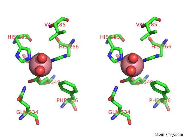
Stereo pair view
Mono view
Stereo pair view
A full contact list of Cobalt with other atoms in the Co binding
site number 1 of Crystal Structure of Human 4-Hydroxyphenylpyruvate Dioxygenase within 5.0Å range:
|
Reference:
E.S.Pilka,
N.Shafqat,
R.Cocking,
J.E.Bray,
T.Krojer,
A.C.W.Pike,
F.Von Delft,
W.W.Yue,
C.H.Arrowsmith,
J.Weigelt,
A.Edwards,
C.Bountra,
U.Oppermann,
K.L.Kavanagh.
Crystal Structure of Human 4-Hydroxyphenylpyruvate Dioxygenase To Be Published.
Page generated: Sun Jul 13 19:02:08 2025
Last articles
Mn in 3NG0Mn in 3NGF
Mn in 3NAI
Mn in 3N4Q
Mn in 3NBP
Mn in 3N5U
Mn in 3N9B
Mn in 3N9D
Mn in 3N39
Mn in 3N3B