Cobalt »
PDB 3mdl-3r61 »
3oqe »
Cobalt in PDB 3oqe: Structure of Opda Mutant Y257F
Enzymatic activity of Structure of Opda Mutant Y257F
All present enzymatic activity of Structure of Opda Mutant Y257F:
3.1.8.1;
3.1.8.1;
Protein crystallography data
The structure of Structure of Opda Mutant Y257F, PDB code: 3oqe
was solved by
F.Ely,
L.W.Guddat,
D.L.Ollis,
G.Schenk,
with X-Ray Crystallography technique. A brief refinement statistics is given in the table below:
| Resolution Low / High (Å) | 21.67 / 1.90 |
| Space group | P 31 2 1 |
| Cell size a, b, c (Å), α, β, γ (°) | 109.048, 109.048, 62.231, 90.00, 90.00, 120.00 |
| R / Rfree (%) | 17.6 / 22.8 |
Cobalt Binding Sites:
The binding sites of Cobalt atom in the Structure of Opda Mutant Y257F
(pdb code 3oqe). This binding sites where shown within
5.0 Angstroms radius around Cobalt atom.
In total 2 binding sites of Cobalt where determined in the Structure of Opda Mutant Y257F, PDB code: 3oqe:
Jump to Cobalt binding site number: 1; 2;
In total 2 binding sites of Cobalt where determined in the Structure of Opda Mutant Y257F, PDB code: 3oqe:
Jump to Cobalt binding site number: 1; 2;
Cobalt binding site 1 out of 2 in 3oqe
Go back to
Cobalt binding site 1 out
of 2 in the Structure of Opda Mutant Y257F
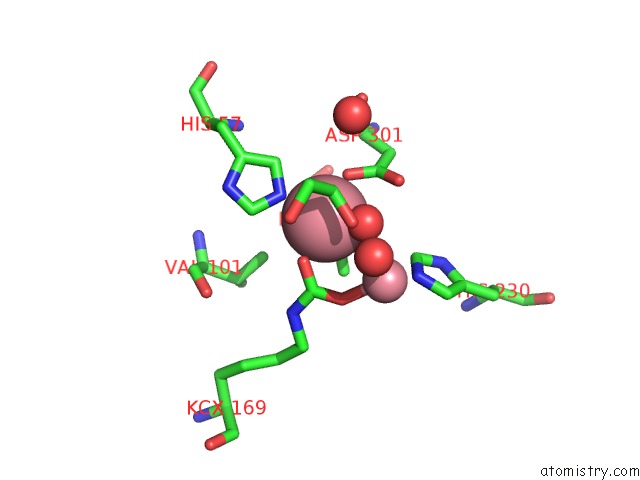
Mono view
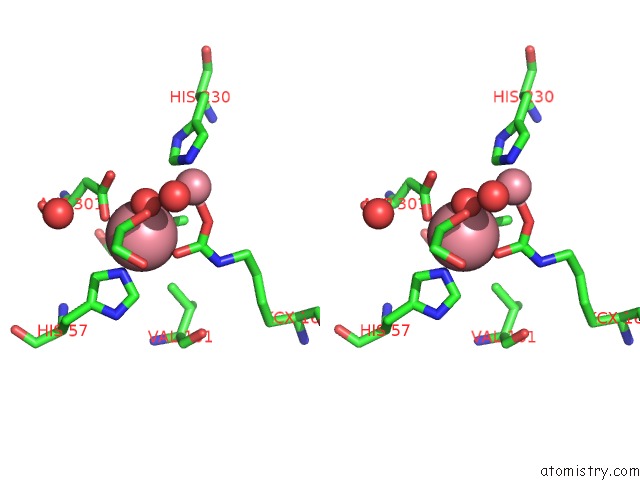
Stereo pair view
Mono view
Stereo pair view
A full contact list of Cobalt with other atoms in the Co binding
site number 1 of Structure of Opda Mutant Y257F within 5.0Å range:
|
Cobalt binding site 2 out of 2 in 3oqe
Go back to
Cobalt binding site 2 out
of 2 in the Structure of Opda Mutant Y257F
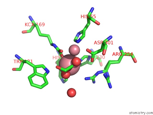
Mono view
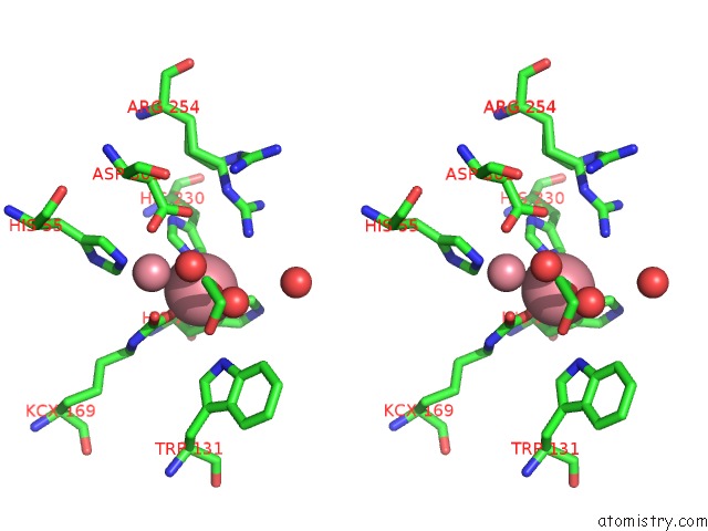
Stereo pair view
Mono view
Stereo pair view
A full contact list of Cobalt with other atoms in the Co binding
site number 2 of Structure of Opda Mutant Y257F within 5.0Å range:
|
Reference:
F.Ely,
K.S.Hadler,
L.R.Gahan,
L.W.Guddat,
D.L.Ollis,
G.Schenk.
The Organophosphate-Degrading Enzyme From Agrobacterium Radiobacter Displays Mechanistic Flexibility For Catalysis. Biochem.J. V. 432 565 2010.
ISSN: ISSN 0264-6021
PubMed: 20868365
DOI: 10.1042/BJ20101054
Page generated: Sun Jul 13 19:14:32 2025
ISSN: ISSN 0264-6021
PubMed: 20868365
DOI: 10.1042/BJ20101054
Last articles
Fe in 2YXOFe in 2YRS
Fe in 2YXC
Fe in 2YNM
Fe in 2YVJ
Fe in 2YP1
Fe in 2YU2
Fe in 2YU1
Fe in 2YQB
Fe in 2YOO