Cobalt »
PDB 3r86-3ura »
3req »
Cobalt in PDB 3req: Methylmalonyl-Coa Mutase, Substrate-Free State (Poor Quality Structure)
Enzymatic activity of Methylmalonyl-Coa Mutase, Substrate-Free State (Poor Quality Structure)
All present enzymatic activity of Methylmalonyl-Coa Mutase, Substrate-Free State (Poor Quality Structure):
5.4.99.2;
5.4.99.2;
Protein crystallography data
The structure of Methylmalonyl-Coa Mutase, Substrate-Free State (Poor Quality Structure), PDB code: 3req
was solved by
P.R.Evans,
F.Mancia,
with X-Ray Crystallography technique. A brief refinement statistics is given in the table below:
| Resolution Low / High (Å) | 20.00 / 2.70 |
| Space group | P 41 21 2 |
| Cell size a, b, c (Å), α, β, γ (°) | 110.910, 110.910, 257.740, 90.00, 90.00, 90.00 |
| R / Rfree (%) | 31.3 / 39.3 |
Cobalt Binding Sites:
The binding sites of Cobalt atom in the Methylmalonyl-Coa Mutase, Substrate-Free State (Poor Quality Structure)
(pdb code 3req). This binding sites where shown within
5.0 Angstroms radius around Cobalt atom.
In total only one binding site of Cobalt was determined in the Methylmalonyl-Coa Mutase, Substrate-Free State (Poor Quality Structure), PDB code: 3req:
In total only one binding site of Cobalt was determined in the Methylmalonyl-Coa Mutase, Substrate-Free State (Poor Quality Structure), PDB code: 3req:
Cobalt binding site 1 out of 1 in 3req
Go back to
Cobalt binding site 1 out
of 1 in the Methylmalonyl-Coa Mutase, Substrate-Free State (Poor Quality Structure)
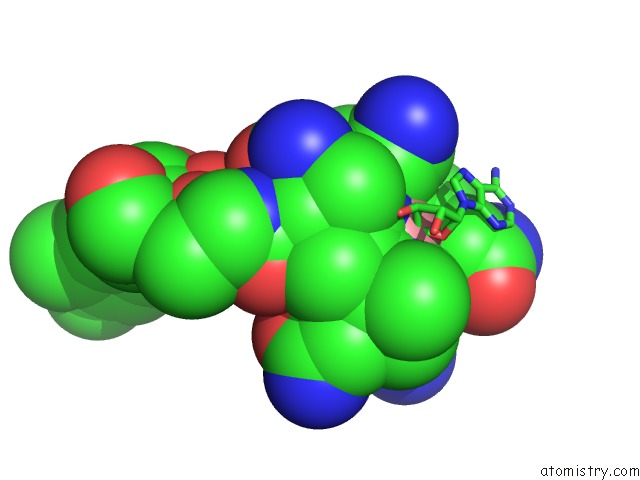
Mono view
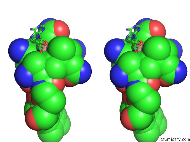
Stereo pair view
Mono view
Stereo pair view
A full contact list of Cobalt with other atoms in the Co binding
site number 1 of Methylmalonyl-Coa Mutase, Substrate-Free State (Poor Quality Structure) within 5.0Å range:
|
Reference:
F.Mancia,
P.R.Evans.
Conformational Changes on Substrate Binding to Methylmalonyl Coa Mutase and New Insights Into the Free Radical Mechanism. Structure V. 6 711 1998.
ISSN: ISSN 0969-2126
PubMed: 9655823
DOI: 10.1016/S0969-2126(98)00073-2
Page generated: Sun Jul 13 19:18:54 2025
ISSN: ISSN 0969-2126
PubMed: 9655823
DOI: 10.1016/S0969-2126(98)00073-2
Last articles
Mg in 9EXRMg in 9EXS
Mg in 9EXQ
Mg in 9EXP
Mg in 9EXO
Mg in 9EX7
Mg in 9EX9
Mg in 9EWJ
Mg in 9EWZ
Mg in 9EW4