Cobalt »
PDB 3r86-3ura »
3rgt »
Cobalt in PDB 3rgt: Crystal Structure of D-Mannonate Dehydratase From Chromohalobacter Salexigens Complexed with D-Arabinohydroxamate
Enzymatic activity of Crystal Structure of D-Mannonate Dehydratase From Chromohalobacter Salexigens Complexed with D-Arabinohydroxamate
All present enzymatic activity of Crystal Structure of D-Mannonate Dehydratase From Chromohalobacter Salexigens Complexed with D-Arabinohydroxamate:
4.2.1.8;
4.2.1.8;
Protein crystallography data
The structure of Crystal Structure of D-Mannonate Dehydratase From Chromohalobacter Salexigens Complexed with D-Arabinohydroxamate, PDB code: 3rgt
was solved by
A.A.Fedorov,
E.V.Fedorov,
D.Wichelecki,
J.A.Gerlt,
S.C.Almo,
with X-Ray Crystallography technique. A brief refinement statistics is given in the table below:
| Resolution Low / High (Å) | 39.85 / 1.90 |
| Space group | P 21 21 2 |
| Cell size a, b, c (Å), α, β, γ (°) | 110.712, 179.657, 85.422, 90.00, 90.00, 90.00 |
| R / Rfree (%) | 17.8 / 22.2 |
Cobalt Binding Sites:
The binding sites of Cobalt atom in the Crystal Structure of D-Mannonate Dehydratase From Chromohalobacter Salexigens Complexed with D-Arabinohydroxamate
(pdb code 3rgt). This binding sites where shown within
5.0 Angstroms radius around Cobalt atom.
In total 4 binding sites of Cobalt where determined in the Crystal Structure of D-Mannonate Dehydratase From Chromohalobacter Salexigens Complexed with D-Arabinohydroxamate, PDB code: 3rgt:
Jump to Cobalt binding site number: 1; 2; 3; 4;
In total 4 binding sites of Cobalt where determined in the Crystal Structure of D-Mannonate Dehydratase From Chromohalobacter Salexigens Complexed with D-Arabinohydroxamate, PDB code: 3rgt:
Jump to Cobalt binding site number: 1; 2; 3; 4;
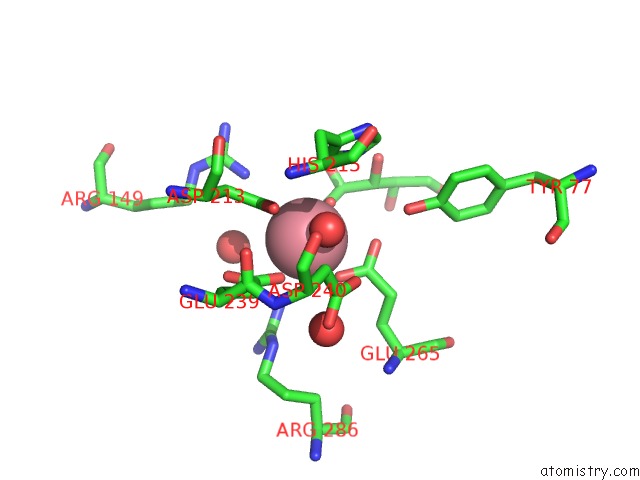
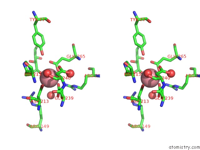
Cobalt binding site 1 out of 4 in 3rgt
Go back to
Cobalt binding site 1 out
of 4 in the Crystal Structure of D-Mannonate Dehydratase From Chromohalobacter Salexigens Complexed with D-Arabinohydroxamate
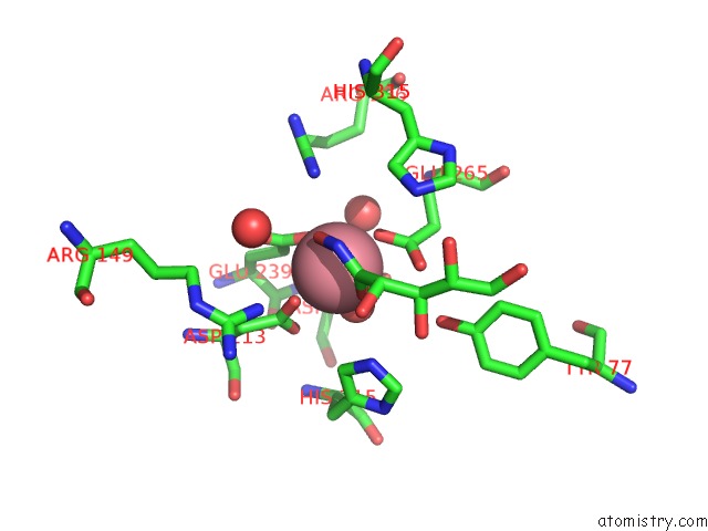
Mono view
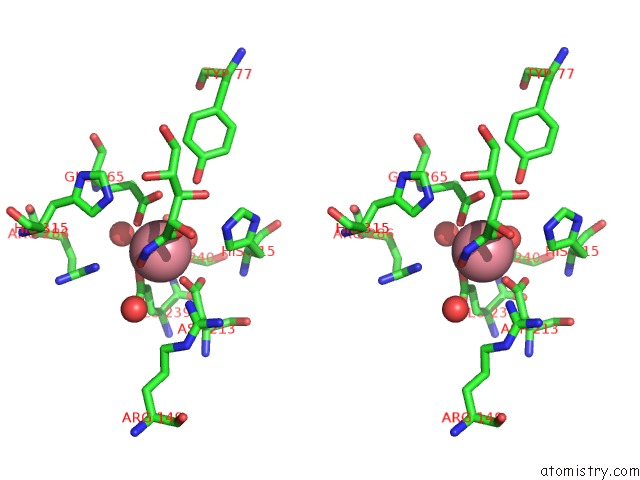
Stereo pair view
Mono view
Stereo pair view
A full contact list of Cobalt with other atoms in the Co binding
site number 1 of Crystal Structure of D-Mannonate Dehydratase From Chromohalobacter Salexigens Complexed with D-Arabinohydroxamate within 5.0Å range:
|
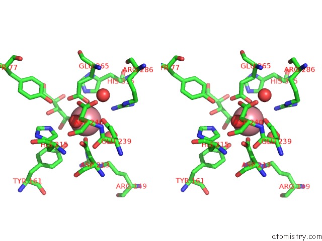
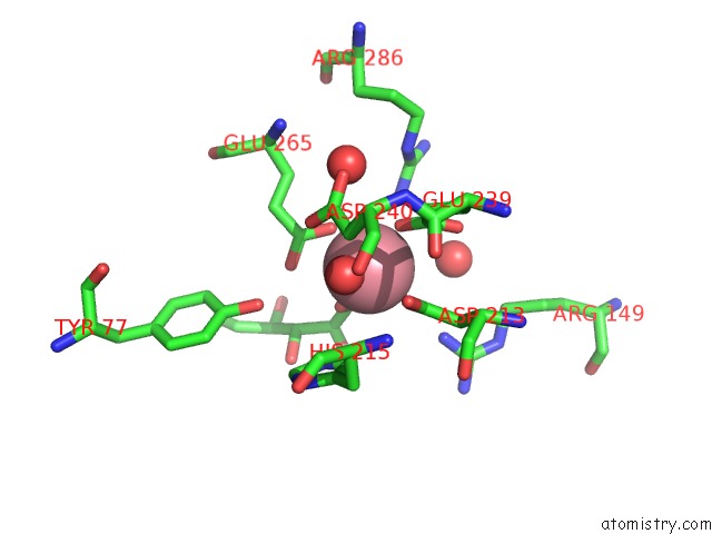
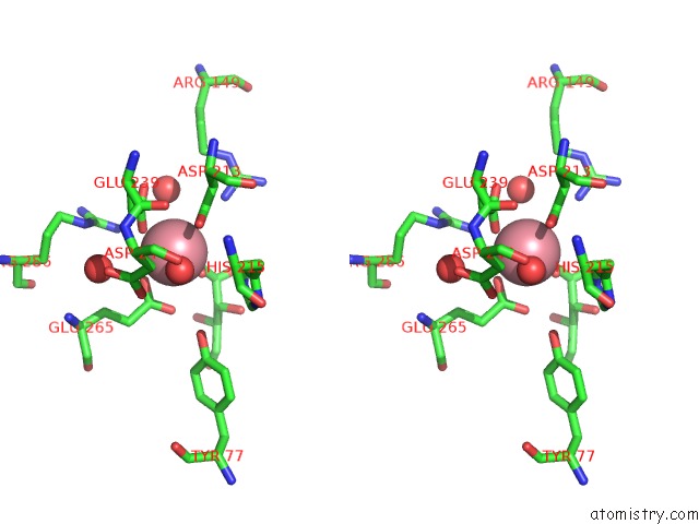
Cobalt binding site 2 out of 4 in 3rgt
Go back to
Cobalt binding site 2 out
of 4 in the Crystal Structure of D-Mannonate Dehydratase From Chromohalobacter Salexigens Complexed with D-Arabinohydroxamate
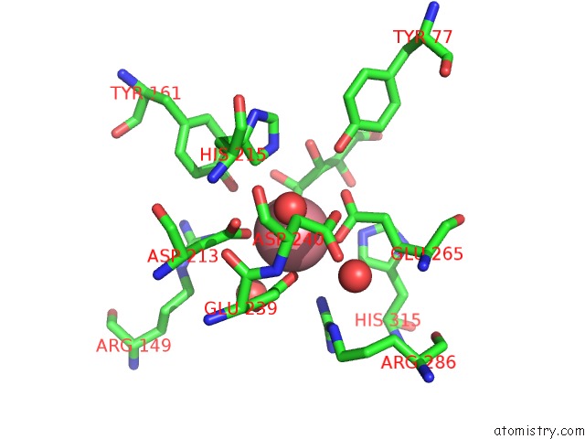
Mono view
Stereo pair view
Mono view
Stereo pair view
A full contact list of Cobalt with other atoms in the Co binding
site number 2 of Crystal Structure of D-Mannonate Dehydratase From Chromohalobacter Salexigens Complexed with D-Arabinohydroxamate within 5.0Å range:
|
Cobalt binding site 3 out of 4 in 3rgt
Go back to
Cobalt binding site 3 out
of 4 in the Crystal Structure of D-Mannonate Dehydratase From Chromohalobacter Salexigens Complexed with D-Arabinohydroxamate
Mono view
Stereo pair view
Mono view
Stereo pair view
A full contact list of Cobalt with other atoms in the Co binding
site number 3 of Crystal Structure of D-Mannonate Dehydratase From Chromohalobacter Salexigens Complexed with D-Arabinohydroxamate within 5.0Å range:
|
Cobalt binding site 4 out of 4 in 3rgt
Go back to
Cobalt binding site 4 out
of 4 in the Crystal Structure of D-Mannonate Dehydratase From Chromohalobacter Salexigens Complexed with D-Arabinohydroxamate
Mono view
Stereo pair view
Mono view
Stereo pair view
A full contact list of Cobalt with other atoms in the Co binding
site number 4 of Crystal Structure of D-Mannonate Dehydratase From Chromohalobacter Salexigens Complexed with D-Arabinohydroxamate within 5.0Å range:
|
Reference:
A.A.Fedorov,
E.V.Fedorov,
D.Wichelecki,
J.A.Gerlt,
S.C.Almo.
Crystal Structure of D-Mannonate Dehydratase From Chromohalobacter Salexigens Complexed with D-Arabinohydroxamate To Be Published.
Page generated: Sun Jul 13 19:18:54 2025
Last articles
Mg in 9F0ZMg in 9F0Y
Mg in 9F07
Mg in 9F0H
Mg in 9EZY
Mg in 9EZD
Mg in 9EXR
Mg in 9EXS
Mg in 9EXQ
Mg in 9EXP