Cobalt »
PDB 3r86-3ura »
3tzi »
Cobalt in PDB 3tzi: X-Ray Crystal Structure of Arachidonic Acid Bound in the Cyclooxygenase Channel of G533V Murine Cox-2
Enzymatic activity of X-Ray Crystal Structure of Arachidonic Acid Bound in the Cyclooxygenase Channel of G533V Murine Cox-2
All present enzymatic activity of X-Ray Crystal Structure of Arachidonic Acid Bound in the Cyclooxygenase Channel of G533V Murine Cox-2:
1.14.99.1;
1.14.99.1;
Protein crystallography data
The structure of X-Ray Crystal Structure of Arachidonic Acid Bound in the Cyclooxygenase Channel of G533V Murine Cox-2, PDB code: 3tzi
was solved by
A.J.Vecchio,
M.G.Malkowski,
with X-Ray Crystallography technique. A brief refinement statistics is given in the table below:
| Resolution Low / High (Å) | 19.99 / 2.15 |
| Space group | I 2 2 2 |
| Cell size a, b, c (Å), α, β, γ (°) | 122.265, 133.508, 181.093, 90.00, 90.00, 90.00 |
| R / Rfree (%) | 17 / 21 |
Cobalt Binding Sites:
The binding sites of Cobalt atom in the X-Ray Crystal Structure of Arachidonic Acid Bound in the Cyclooxygenase Channel of G533V Murine Cox-2
(pdb code 3tzi). This binding sites where shown within
5.0 Angstroms radius around Cobalt atom.
In total 2 binding sites of Cobalt where determined in the X-Ray Crystal Structure of Arachidonic Acid Bound in the Cyclooxygenase Channel of G533V Murine Cox-2, PDB code: 3tzi:
Jump to Cobalt binding site number: 1; 2;
In total 2 binding sites of Cobalt where determined in the X-Ray Crystal Structure of Arachidonic Acid Bound in the Cyclooxygenase Channel of G533V Murine Cox-2, PDB code: 3tzi:
Jump to Cobalt binding site number: 1; 2;
Cobalt binding site 1 out of 2 in 3tzi
Go back to
Cobalt binding site 1 out
of 2 in the X-Ray Crystal Structure of Arachidonic Acid Bound in the Cyclooxygenase Channel of G533V Murine Cox-2
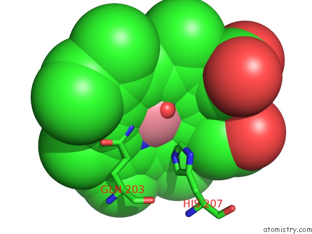
Mono view
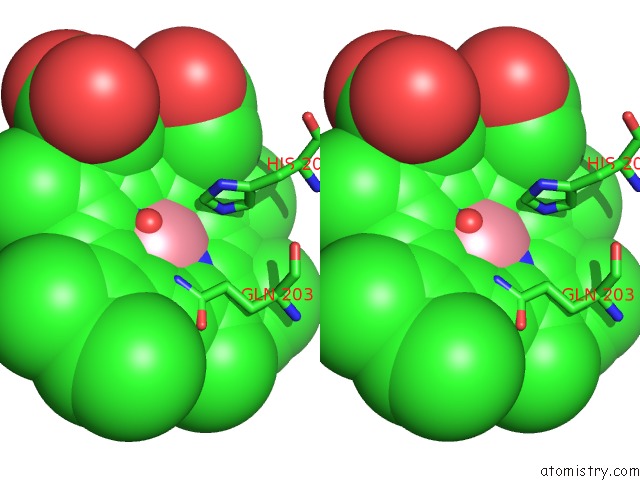
Stereo pair view
Mono view
Stereo pair view
A full contact list of Cobalt with other atoms in the Co binding
site number 1 of X-Ray Crystal Structure of Arachidonic Acid Bound in the Cyclooxygenase Channel of G533V Murine Cox-2 within 5.0Å range:
|
Cobalt binding site 2 out of 2 in 3tzi
Go back to
Cobalt binding site 2 out
of 2 in the X-Ray Crystal Structure of Arachidonic Acid Bound in the Cyclooxygenase Channel of G533V Murine Cox-2
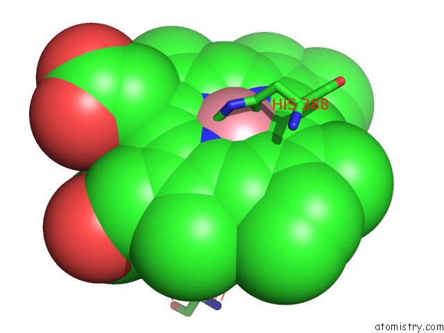
Mono view
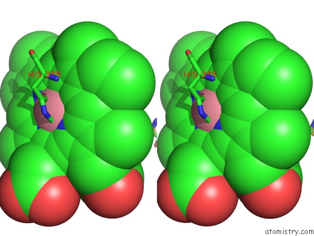
Stereo pair view
Mono view
Stereo pair view
A full contact list of Cobalt with other atoms in the Co binding
site number 2 of X-Ray Crystal Structure of Arachidonic Acid Bound in the Cyclooxygenase Channel of G533V Murine Cox-2 within 5.0Å range:
|
Reference:
A.J.Vecchio,
B.J.Orlando,
R.Nandagiri,
M.G.Malkowski.
Investigating Substrate Promiscuity in Cyclooxygenase-2: the Role of Arg-120 and Residues Lining the Hydrophobic Groove. J.Biol.Chem. V. 287 24619 2012.
ISSN: ISSN 0021-9258
PubMed: 22637474
DOI: 10.1074/JBC.M112.372243
Page generated: Sun Jul 13 19:27:00 2025
ISSN: ISSN 0021-9258
PubMed: 22637474
DOI: 10.1074/JBC.M112.372243
Last articles
Mg in 9F28Mg in 9F2R
Mg in 9F12
Mg in 9F11
Mg in 9F10
Mg in 9F0Z
Mg in 9F0Y
Mg in 9F07
Mg in 9F0H
Mg in 9EZY