Cobalt »
PDB 3urb-3w71 »
3vyh »
Cobalt in PDB 3vyh: Crystal Structure of AW116R Mutant of Nitrile Hydratase From Pseudonocardia Thermophilla
Enzymatic activity of Crystal Structure of AW116R Mutant of Nitrile Hydratase From Pseudonocardia Thermophilla
All present enzymatic activity of Crystal Structure of AW116R Mutant of Nitrile Hydratase From Pseudonocardia Thermophilla:
4.2.1.84;
4.2.1.84;
Protein crystallography data
The structure of Crystal Structure of AW116R Mutant of Nitrile Hydratase From Pseudonocardia Thermophilla, PDB code: 3vyh
was solved by
Y.Yamanaka,
M.Sato,
T.Arakawa,
S.Namima,
S.Hori,
A.Ohtaki,
K.Noguchi,
Y.Katayama,
M.Yohda,
M.Odaka,
with X-Ray Crystallography technique. A brief refinement statistics is given in the table below:
| Resolution Low / High (Å) | 48.44 / 1.63 |
| Space group | P 32 2 1 |
| Cell size a, b, c (Å), α, β, γ (°) | 65.683, 65.683, 184.796, 90.00, 90.00, 120.00 |
| R / Rfree (%) | 17.4 / 20.3 |
Cobalt Binding Sites:
The binding sites of Cobalt atom in the Crystal Structure of AW116R Mutant of Nitrile Hydratase From Pseudonocardia Thermophilla
(pdb code 3vyh). This binding sites where shown within
5.0 Angstroms radius around Cobalt atom.
In total only one binding site of Cobalt was determined in the Crystal Structure of AW116R Mutant of Nitrile Hydratase From Pseudonocardia Thermophilla, PDB code: 3vyh:
In total only one binding site of Cobalt was determined in the Crystal Structure of AW116R Mutant of Nitrile Hydratase From Pseudonocardia Thermophilla, PDB code: 3vyh:
Cobalt binding site 1 out of 1 in 3vyh
Go back to
Cobalt binding site 1 out
of 1 in the Crystal Structure of AW116R Mutant of Nitrile Hydratase From Pseudonocardia Thermophilla
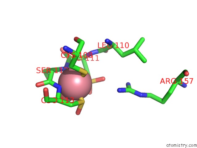
Mono view
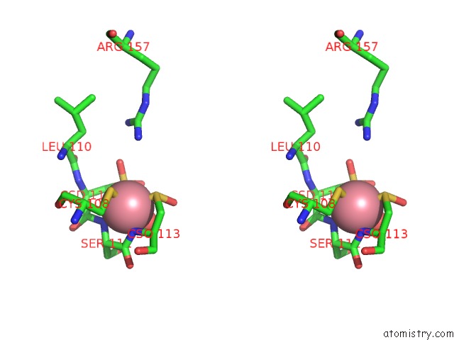
Stereo pair view
Mono view
Stereo pair view
A full contact list of Cobalt with other atoms in the Co binding
site number 1 of Crystal Structure of AW116R Mutant of Nitrile Hydratase From Pseudonocardia Thermophilla within 5.0Å range:
|
Reference:
Y.Yamanaka,
M.Sato,
T.Arakawa,
S.Namima,
S.Hori,
A.Ohtaki,
K.Noguchi,
Y.Katayama,
M.Yohda,
M.Odaka.
Effects of Argnine Residue Around the Substrate Pocket on the Substrate Specificity of Thiocyanate Hydrolase To Be Published.
Page generated: Sun Jul 13 19:31:46 2025
Last articles
Mg in 6I4FMg in 6I4I
Mg in 6I1L
Mg in 6I4D
Mg in 6I4E
Mg in 6I3D
Mg in 6I3C
Mg in 6I36
Mg in 6I2U
Mg in 6I39