Cobalt »
PDB 4as7-4fck »
4etl »
Cobalt in PDB 4etl: Crystallographic Structure of Phenylalanine Hydroxylase From Chromobacterium Violaceum F258A Mutation
Enzymatic activity of Crystallographic Structure of Phenylalanine Hydroxylase From Chromobacterium Violaceum F258A Mutation
All present enzymatic activity of Crystallographic Structure of Phenylalanine Hydroxylase From Chromobacterium Violaceum F258A Mutation:
1.14.16.1;
1.14.16.1;
Protein crystallography data
The structure of Crystallographic Structure of Phenylalanine Hydroxylase From Chromobacterium Violaceum F258A Mutation, PDB code: 4etl
was solved by
J.A.Ronau,
L.P.Paul,
I.R.Corn,
K.T.Wagner,
M.M.Abu-Omar,
C.Das,
with X-Ray Crystallography technique. A brief refinement statistics is given in the table below:
| Resolution Low / High (Å) | 22.68 / 1.49 |
| Space group | P 1 |
| Cell size a, b, c (Å), α, β, γ (°) | 36.780, 38.553, 48.044, 76.47, 73.00, 85.36 |
| R / Rfree (%) | 16.5 / 21.4 |
Cobalt Binding Sites:
The binding sites of Cobalt atom in the Crystallographic Structure of Phenylalanine Hydroxylase From Chromobacterium Violaceum F258A Mutation
(pdb code 4etl). This binding sites where shown within
5.0 Angstroms radius around Cobalt atom.
In total 2 binding sites of Cobalt where determined in the Crystallographic Structure of Phenylalanine Hydroxylase From Chromobacterium Violaceum F258A Mutation, PDB code: 4etl:
Jump to Cobalt binding site number: 1; 2;
In total 2 binding sites of Cobalt where determined in the Crystallographic Structure of Phenylalanine Hydroxylase From Chromobacterium Violaceum F258A Mutation, PDB code: 4etl:
Jump to Cobalt binding site number: 1; 2;
Cobalt binding site 1 out of 2 in 4etl
Go back to
Cobalt binding site 1 out
of 2 in the Crystallographic Structure of Phenylalanine Hydroxylase From Chromobacterium Violaceum F258A Mutation
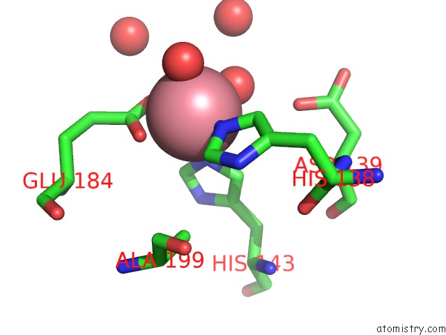
Mono view
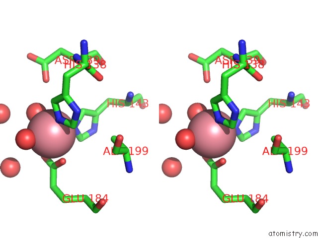
Stereo pair view
Mono view
Stereo pair view
A full contact list of Cobalt with other atoms in the Co binding
site number 1 of Crystallographic Structure of Phenylalanine Hydroxylase From Chromobacterium Violaceum F258A Mutation within 5.0Å range:
|
Cobalt binding site 2 out of 2 in 4etl
Go back to
Cobalt binding site 2 out
of 2 in the Crystallographic Structure of Phenylalanine Hydroxylase From Chromobacterium Violaceum F258A Mutation
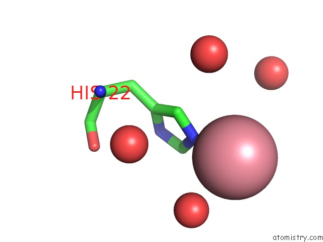
Mono view
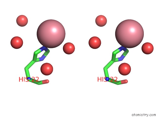
Stereo pair view
Mono view
Stereo pair view
A full contact list of Cobalt with other atoms in the Co binding
site number 2 of Crystallographic Structure of Phenylalanine Hydroxylase From Chromobacterium Violaceum F258A Mutation within 5.0Å range:
|
Reference:
J.A.Ronau,
L.N.Paul,
J.E.Fuchs,
I.R.Corn,
K.T.Wagner,
K.R.Liedl,
M.M.Abu-Omar,
C.Das.
An Additional Substrate Binding Site in A Bacterial Phenylalanine Hydroxylase. Eur.Biophys.J. V. 42 691 2013.
ISSN: ISSN 0175-7571
PubMed: 23860686
DOI: 10.1007/S00249-013-0919-8
Page generated: Sun Jul 13 19:42:13 2025
ISSN: ISSN 0175-7571
PubMed: 23860686
DOI: 10.1007/S00249-013-0919-8
Last articles
Mg in 9EXRMg in 9EXS
Mg in 9EXQ
Mg in 9EXP
Mg in 9EXO
Mg in 9EX7
Mg in 9EX9
Mg in 9EWJ
Mg in 9EWZ
Mg in 9EW4