Cobalt »
PDB 4fe5-4j0b »
4hcq »
Cobalt in PDB 4hcq: Crystal Structure of Glmu From Mycobacterium Tuberculosis in Complex with Glucosamine-1-Phosphate
Enzymatic activity of Crystal Structure of Glmu From Mycobacterium Tuberculosis in Complex with Glucosamine-1-Phosphate
All present enzymatic activity of Crystal Structure of Glmu From Mycobacterium Tuberculosis in Complex with Glucosamine-1-Phosphate:
2.3.1.157; 2.7.7.23;
2.3.1.157; 2.7.7.23;
Protein crystallography data
The structure of Crystal Structure of Glmu From Mycobacterium Tuberculosis in Complex with Glucosamine-1-Phosphate, PDB code: 4hcq
was solved by
P.K.A.Jagtap,
S.K.Verma,
N.Vithani,
with X-Ray Crystallography technique. A brief refinement statistics is given in the table below:
| Resolution Low / High (Å) | 19.93 / 2.60 |
| Space group | H 3 |
| Cell size a, b, c (Å), α, β, γ (°) | 79.270, 79.270, 276.730, 90.00, 90.00, 120.00 |
| R / Rfree (%) | 19.9 / 26.5 |
Other elements in 4hcq:
The structure of Crystal Structure of Glmu From Mycobacterium Tuberculosis in Complex with Glucosamine-1-Phosphate also contains other interesting chemical elements:
| Magnesium | (Mg) | 2 atoms |
Cobalt Binding Sites:
The binding sites of Cobalt atom in the Crystal Structure of Glmu From Mycobacterium Tuberculosis in Complex with Glucosamine-1-Phosphate
(pdb code 4hcq). This binding sites where shown within
5.0 Angstroms radius around Cobalt atom.
In total only one binding site of Cobalt was determined in the Crystal Structure of Glmu From Mycobacterium Tuberculosis in Complex with Glucosamine-1-Phosphate, PDB code: 4hcq:
In total only one binding site of Cobalt was determined in the Crystal Structure of Glmu From Mycobacterium Tuberculosis in Complex with Glucosamine-1-Phosphate, PDB code: 4hcq:
Cobalt binding site 1 out of 1 in 4hcq
Go back to
Cobalt binding site 1 out
of 1 in the Crystal Structure of Glmu From Mycobacterium Tuberculosis in Complex with Glucosamine-1-Phosphate
Mono view
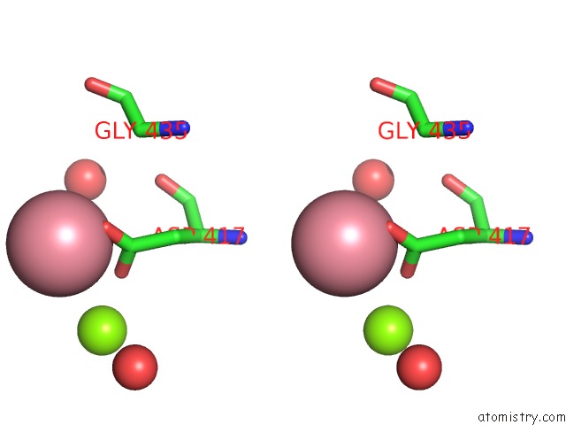
Stereo pair view
Mono view
Stereo pair view
A full contact list of Cobalt with other atoms in the Co binding
site number 1 of Crystal Structure of Glmu From Mycobacterium Tuberculosis in Complex with Glucosamine-1-Phosphate within 5.0Å range:
|
Reference:
P.K.A.Jagtap,
S.K.Verma,
N.Vithani,
V.S.Bais,
B.Prakash.
Crystal Structures Identify An Atypical Two-Metal-Ion Mechanism For Uridyltransfer in Glmu: Its Significance to Sugar Nucleotidyl Transferases J.Mol.Biol. V. 425 1745 2013.
ISSN: ISSN 0022-2836
PubMed: 23485416
DOI: 10.1016/J.JMB.2013.02.019
Page generated: Sun Jul 13 19:47:24 2025
ISSN: ISSN 0022-2836
PubMed: 23485416
DOI: 10.1016/J.JMB.2013.02.019
Last articles
Mg in 4DR2Mg in 4DR3
Mg in 4DR1
Mg in 4DPG
Mg in 4DQP
Mg in 4DQQ
Mg in 4DPM
Mg in 4DPV
Mg in 4DQI
Mg in 4DOB