Cobalt »
PDB 4fe5-4j0b »
4hei »
Cobalt in PDB 4hei: 2A X-Ray Structure of Hpf From Vibrio Cholerae
Protein crystallography data
The structure of 2A X-Ray Structure of Hpf From Vibrio Cholerae, PDB code: 4hei
was solved by
H.De Bari,
E.A.Berry,
with X-Ray Crystallography technique. A brief refinement statistics is given in the table below:
| Resolution Low / High (Å) | 43.62 / 1.60 |
| Space group | P 41 21 2 |
| Cell size a, b, c (Å), α, β, γ (°) | 46.354, 46.354, 174.492, 90.00, 90.00, 90.00 |
| R / Rfree (%) | 26.2 / 28.1 |
Cobalt Binding Sites:
The binding sites of Cobalt atom in the 2A X-Ray Structure of Hpf From Vibrio Cholerae
(pdb code 4hei). This binding sites where shown within
5.0 Angstroms radius around Cobalt atom.
In total 4 binding sites of Cobalt where determined in the 2A X-Ray Structure of Hpf From Vibrio Cholerae, PDB code: 4hei:
Jump to Cobalt binding site number: 1; 2; 3; 4;
In total 4 binding sites of Cobalt where determined in the 2A X-Ray Structure of Hpf From Vibrio Cholerae, PDB code: 4hei:
Jump to Cobalt binding site number: 1; 2; 3; 4;
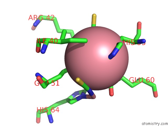
Cobalt binding site 1 out of 4 in 4hei
Go back to
Cobalt binding site 1 out
of 4 in the 2A X-Ray Structure of Hpf From Vibrio Cholerae
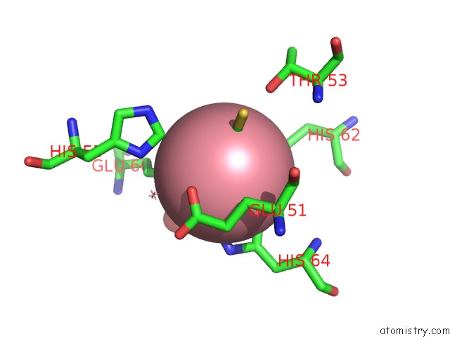
Mono view
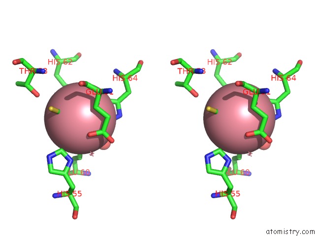
Stereo pair view
Mono view
Stereo pair view
A full contact list of Cobalt with other atoms in the Co binding
site number 1 of 2A X-Ray Structure of Hpf From Vibrio Cholerae within 5.0Å range:
|
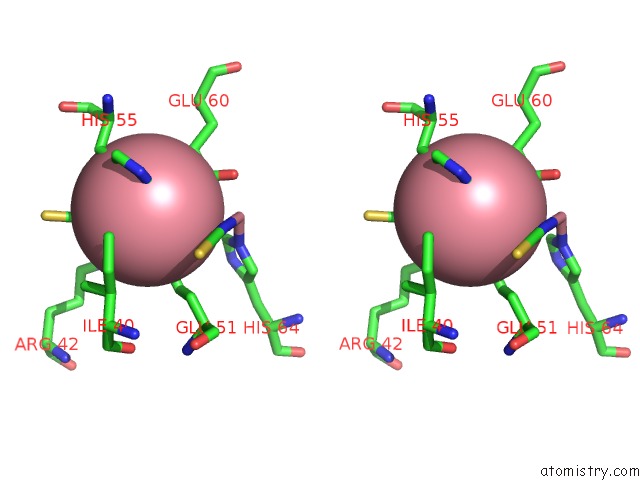
Cobalt binding site 2 out of 4 in 4hei
Go back to
Cobalt binding site 2 out
of 4 in the 2A X-Ray Structure of Hpf From Vibrio Cholerae
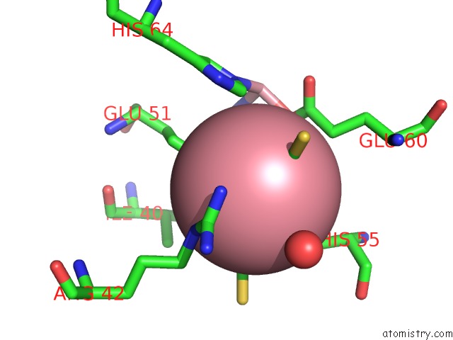
Mono view
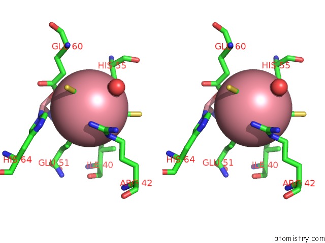
Stereo pair view
Mono view
Stereo pair view
A full contact list of Cobalt with other atoms in the Co binding
site number 2 of 2A X-Ray Structure of Hpf From Vibrio Cholerae within 5.0Å range:
|
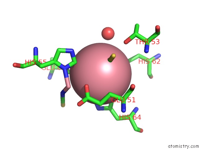
Cobalt binding site 3 out of 4 in 4hei
Go back to
Cobalt binding site 3 out
of 4 in the 2A X-Ray Structure of Hpf From Vibrio Cholerae
Mono view
Stereo pair view
Mono view
Stereo pair view
A full contact list of Cobalt with other atoms in the Co binding
site number 3 of 2A X-Ray Structure of Hpf From Vibrio Cholerae within 5.0Å range:
|
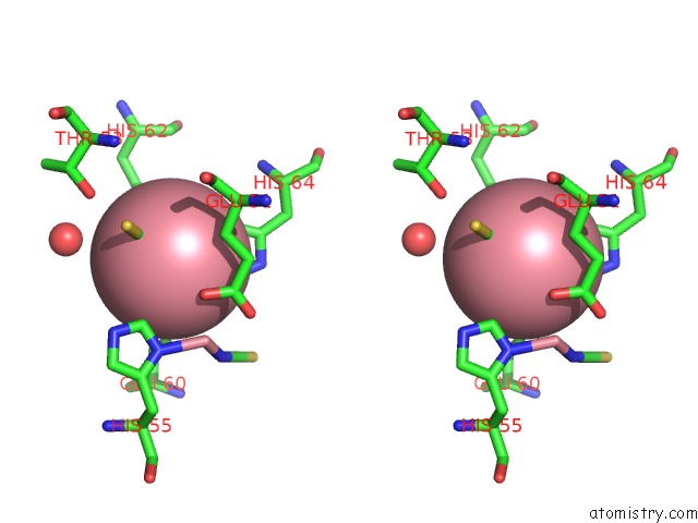
Cobalt binding site 4 out of 4 in 4hei
Go back to
Cobalt binding site 4 out
of 4 in the 2A X-Ray Structure of Hpf From Vibrio Cholerae
Mono view
Stereo pair view
Mono view
Stereo pair view
A full contact list of Cobalt with other atoms in the Co binding
site number 4 of 2A X-Ray Structure of Hpf From Vibrio Cholerae within 5.0Å range:
|
Reference:
H.De Bari,
E.A.Berry.
Structure of Vibrio Cholerae Ribosome Hibernation Promoting Factor. Acta Crystallogr.,Sect.F V. 69 228 2013.
ISSN: ESSN 1744-3091
PubMed: 23519794
DOI: 10.1107/S1744309113000961
Page generated: Sun Jul 13 19:47:26 2025
ISSN: ESSN 1744-3091
PubMed: 23519794
DOI: 10.1107/S1744309113000961
Last articles
Mg in 4DR3Mg in 4DR1
Mg in 4DPG
Mg in 4DQP
Mg in 4DQQ
Mg in 4DPM
Mg in 4DPV
Mg in 4DQI
Mg in 4DOB
Mg in 4DOC