Cobalt »
PDB 4j2m-4ngl »
4mag »
Cobalt in PDB 4mag: Crystal Structure of the Periplasmic Sialic Acid Binding Protein From Vibrio Cholerea
Protein crystallography data
The structure of Crystal Structure of the Periplasmic Sialic Acid Binding Protein From Vibrio Cholerea, PDB code: 4mag
was solved by
S.Ramaswamy,
C.Cho,
M.A.Apicella,
with X-Ray Crystallography technique. A brief refinement statistics is given in the table below:
| Resolution Low / High (Å) | 11.73 / 1.45 |
| Space group | I 2 2 2 |
| Cell size a, b, c (Å), α, β, γ (°) | 74.977, 82.315, 119.354, 90.00, 90.00, 90.00 |
| R / Rfree (%) | 18.2 / 21.9 |
Cobalt Binding Sites:
The binding sites of Cobalt atom in the Crystal Structure of the Periplasmic Sialic Acid Binding Protein From Vibrio Cholerea
(pdb code 4mag). This binding sites where shown within
5.0 Angstroms radius around Cobalt atom.
In total only one binding site of Cobalt was determined in the Crystal Structure of the Periplasmic Sialic Acid Binding Protein From Vibrio Cholerea, PDB code: 4mag:
In total only one binding site of Cobalt was determined in the Crystal Structure of the Periplasmic Sialic Acid Binding Protein From Vibrio Cholerea, PDB code: 4mag:
Cobalt binding site 1 out of 1 in 4mag
Go back to
Cobalt binding site 1 out
of 1 in the Crystal Structure of the Periplasmic Sialic Acid Binding Protein From Vibrio Cholerea
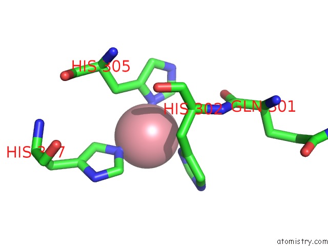
Mono view
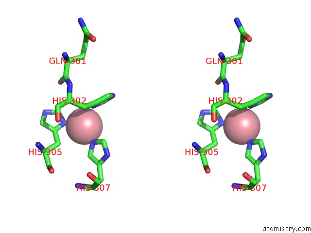
Stereo pair view
Mono view
Stereo pair view
A full contact list of Cobalt with other atoms in the Co binding
site number 1 of Crystal Structure of the Periplasmic Sialic Acid Binding Protein From Vibrio Cholerea within 5.0Å range:
|
Reference:
T.Gangi Setty,
C.Cho,
S.Govindappa,
M.A.Apicella,
S.Ramaswamy.
Bacterial Periplasmic Sialic Acid-Binding Proteins Exhibit A Conserved Binding Site. Acta Crystallogr.,Sect.D V. 70 1801 2014.
ISSN: ISSN 0907-4449
PubMed: 25004958
DOI: 10.1107/S139900471400830X
Page generated: Sun Jul 13 19:56:30 2025
ISSN: ISSN 0907-4449
PubMed: 25004958
DOI: 10.1107/S139900471400830X
Last articles
Mg in 9F28Mg in 9F2R
Mg in 9F12
Mg in 9F11
Mg in 9F10
Mg in 9F0Z
Mg in 9F0Y
Mg in 9F07
Mg in 9F0H
Mg in 9EZY