Cobalt »
PDB 4xim-5d6e »
5cel »
Cobalt in PDB 5cel: CBH1 (E212Q) Cellotetraose Complex
Enzymatic activity of CBH1 (E212Q) Cellotetraose Complex
All present enzymatic activity of CBH1 (E212Q) Cellotetraose Complex:
3.2.1.91;
3.2.1.91;
Protein crystallography data
The structure of CBH1 (E212Q) Cellotetraose Complex, PDB code: 5cel
was solved by
C.Divne,
J.Stahlberg,
T.A.Jones,
with X-Ray Crystallography technique. A brief refinement statistics is given in the table below:
| Resolution Low / High (Å) | 7.50 / 1.90 |
| Space group | I 2 2 2 |
| Cell size a, b, c (Å), α, β, γ (°) | 83.050, 82.950, 110.850, 90.00, 90.00, 90.00 |
| R / Rfree (%) | 19.6 / 24 |
Cobalt Binding Sites:
The binding sites of Cobalt atom in the CBH1 (E212Q) Cellotetraose Complex
(pdb code 5cel). This binding sites where shown within
5.0 Angstroms radius around Cobalt atom.
In total 2 binding sites of Cobalt where determined in the CBH1 (E212Q) Cellotetraose Complex, PDB code: 5cel:
Jump to Cobalt binding site number: 1; 2;
In total 2 binding sites of Cobalt where determined in the CBH1 (E212Q) Cellotetraose Complex, PDB code: 5cel:
Jump to Cobalt binding site number: 1; 2;
Cobalt binding site 1 out of 2 in 5cel
Go back to
Cobalt binding site 1 out
of 2 in the CBH1 (E212Q) Cellotetraose Complex
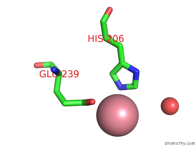
Mono view
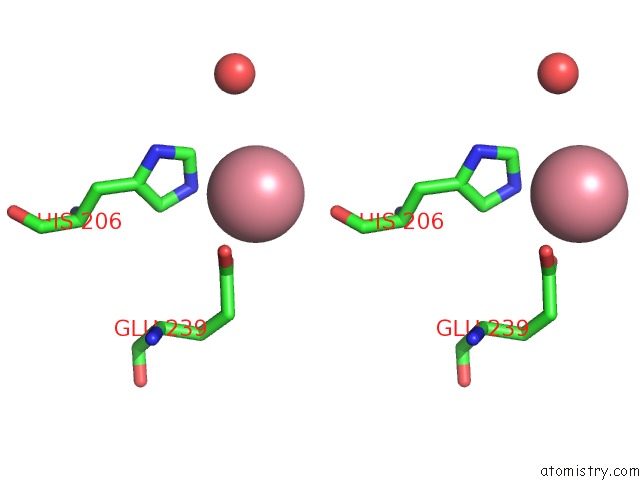
Stereo pair view
Mono view
Stereo pair view
A full contact list of Cobalt with other atoms in the Co binding
site number 1 of CBH1 (E212Q) Cellotetraose Complex within 5.0Å range:
|
Cobalt binding site 2 out of 2 in 5cel
Go back to
Cobalt binding site 2 out
of 2 in the CBH1 (E212Q) Cellotetraose Complex
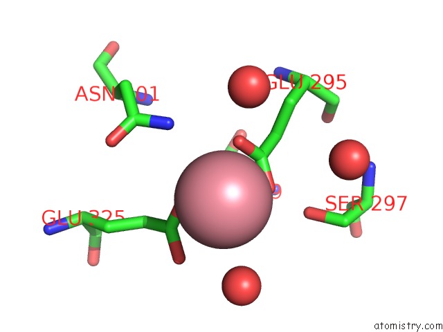
Mono view
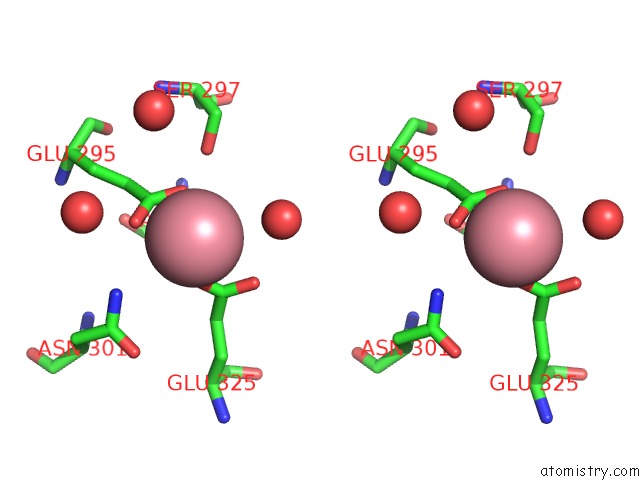
Stereo pair view
Mono view
Stereo pair view
A full contact list of Cobalt with other atoms in the Co binding
site number 2 of CBH1 (E212Q) Cellotetraose Complex within 5.0Å range:
|
Reference:
C.Divne,
J.Stahlberg,
T.T.Teeri,
T.A.Jones.
High-Resolution Crystal Structures Reveal How A Cellulose Chain Is Bound in the 50 A Long Tunnel of Cellobiohydrolase I From Trichoderma Reesei. J.Mol.Biol. V. 275 309 1998.
ISSN: ISSN 0022-2836
PubMed: 9466911
DOI: 10.1006/JMBI.1997.1437
Page generated: Sun Jul 13 20:16:51 2025
ISSN: ISSN 0022-2836
PubMed: 9466911
DOI: 10.1006/JMBI.1997.1437
Last articles
Fe in 2YXOFe in 2YRS
Fe in 2YXC
Fe in 2YNM
Fe in 2YVJ
Fe in 2YP1
Fe in 2YU2
Fe in 2YU1
Fe in 2YQB
Fe in 2YOO