Cobalt »
PDB 5d6f-5ikv »
5f1a »
Cobalt in PDB 5f1a: The Crystal Structure of Salicylate Bound to Human Cyclooxygenase-2
Enzymatic activity of The Crystal Structure of Salicylate Bound to Human Cyclooxygenase-2
All present enzymatic activity of The Crystal Structure of Salicylate Bound to Human Cyclooxygenase-2:
1.14.99.1;
1.14.99.1;
Protein crystallography data
The structure of The Crystal Structure of Salicylate Bound to Human Cyclooxygenase-2, PDB code: 5f1a
was solved by
M.J.Lucido,
B.J.Orlando,
M.G.Malkowski,
with X-Ray Crystallography technique. A brief refinement statistics is given in the table below:
| Resolution Low / High (Å) | 33.32 / 2.38 |
| Space group | I 2 2 2 |
| Cell size a, b, c (Å), α, β, γ (°) | 118.410, 132.660, 178.740, 90.00, 90.00, 90.00 |
| R / Rfree (%) | 17.4 / 21.8 |
Cobalt Binding Sites:
The binding sites of Cobalt atom in the The Crystal Structure of Salicylate Bound to Human Cyclooxygenase-2
(pdb code 5f1a). This binding sites where shown within
5.0 Angstroms radius around Cobalt atom.
In total 2 binding sites of Cobalt where determined in the The Crystal Structure of Salicylate Bound to Human Cyclooxygenase-2, PDB code: 5f1a:
Jump to Cobalt binding site number: 1; 2;
In total 2 binding sites of Cobalt where determined in the The Crystal Structure of Salicylate Bound to Human Cyclooxygenase-2, PDB code: 5f1a:
Jump to Cobalt binding site number: 1; 2;
Cobalt binding site 1 out of 2 in 5f1a
Go back to
Cobalt binding site 1 out
of 2 in the The Crystal Structure of Salicylate Bound to Human Cyclooxygenase-2
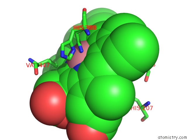
Mono view
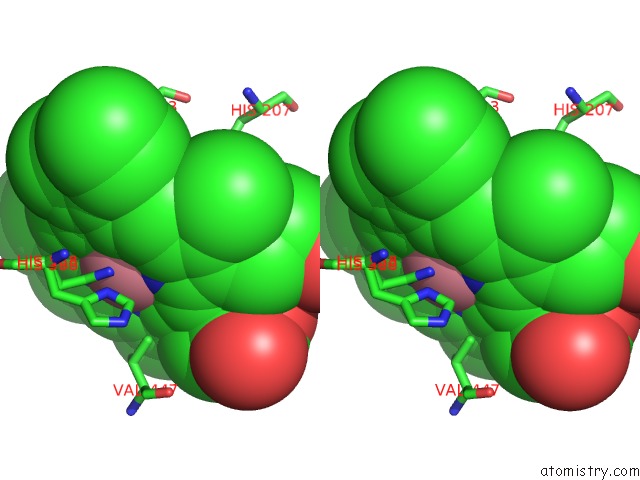
Stereo pair view
Mono view
Stereo pair view
A full contact list of Cobalt with other atoms in the Co binding
site number 1 of The Crystal Structure of Salicylate Bound to Human Cyclooxygenase-2 within 5.0Å range:
|
Cobalt binding site 2 out of 2 in 5f1a
Go back to
Cobalt binding site 2 out
of 2 in the The Crystal Structure of Salicylate Bound to Human Cyclooxygenase-2
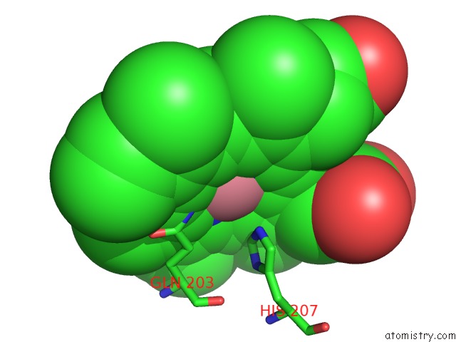
Mono view
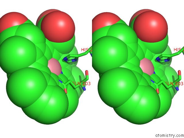
Stereo pair view
Mono view
Stereo pair view
A full contact list of Cobalt with other atoms in the Co binding
site number 2 of The Crystal Structure of Salicylate Bound to Human Cyclooxygenase-2 within 5.0Å range:
|
Reference:
M.J.Lucido,
B.J.Orlando,
A.J.Vecchio,
M.G.Malkowski.
Crystal Structure of Aspirin-Acetylated Human Cyclooxygenase-2: Insight Into the Formation of Products with Reversed Stereochemistry. Biochemistry V. 55 1226 2016.
ISSN: ISSN 0006-2960
PubMed: 26859324
DOI: 10.1021/ACS.BIOCHEM.5B01378
Page generated: Sun Jul 13 20:22:10 2025
ISSN: ISSN 0006-2960
PubMed: 26859324
DOI: 10.1021/ACS.BIOCHEM.5B01378
Last articles
K in 4R33K in 4R2C
K in 4QXG
K in 4QRH
K in 4QNE
K in 4QGC
K in 4QKA
K in 4QE9
K in 4QG8
K in 4QK8