Cobalt »
PDB 5img-5np4 »
5iqn »
Cobalt in PDB 5iqn: Crystal Structure of the E. Coli Type 1 Pilus Subunit Fimg (Engineered Variant with Substitution Q134E; N-Terminal Fimg Residues 1-12 Truncated) in Complex with the Donor Strand Peptide DSF_SRIRIRGYVR
Protein crystallography data
The structure of Crystal Structure of the E. Coli Type 1 Pilus Subunit Fimg (Engineered Variant with Substitution Q134E; N-Terminal Fimg Residues 1-12 Truncated) in Complex with the Donor Strand Peptide DSF_SRIRIRGYVR, PDB code: 5iqn
was solved by
C.Giese,
J.Eras,
A.Kern,
M.A.Scharer,
G.Capitani,
R.Glockshuber,
with X-Ray Crystallography technique. A brief refinement statistics is given in the table below:
| Resolution Low / High (Å) | 25.38 / 1.00 |
| Space group | P 1 21 1 |
| Cell size a, b, c (Å), α, β, γ (°) | 25.680, 52.880, 83.140, 90.00, 98.71, 90.00 |
| R / Rfree (%) | 11.8 / 14.6 |
Cobalt Binding Sites:
The binding sites of Cobalt atom in the Crystal Structure of the E. Coli Type 1 Pilus Subunit Fimg (Engineered Variant with Substitution Q134E; N-Terminal Fimg Residues 1-12 Truncated) in Complex with the Donor Strand Peptide DSF_SRIRIRGYVR
(pdb code 5iqn). This binding sites where shown within
5.0 Angstroms radius around Cobalt atom.
In total 2 binding sites of Cobalt where determined in the Crystal Structure of the E. Coli Type 1 Pilus Subunit Fimg (Engineered Variant with Substitution Q134E; N-Terminal Fimg Residues 1-12 Truncated) in Complex with the Donor Strand Peptide DSF_SRIRIRGYVR, PDB code: 5iqn:
Jump to Cobalt binding site number: 1; 2;
Cobalt binding site 1 out of 2 in 5iqn
Go back to
Cobalt binding site 1 out
of 2 in the Crystal Structure of the E. Coli Type 1 Pilus Subunit Fimg (Engineered Variant with Substitution Q134E; N-Terminal Fimg Residues 1-12 Truncated) in Complex with the Donor Strand Peptide DSF_SRIRIRGYVR
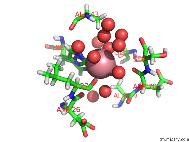
Mono view
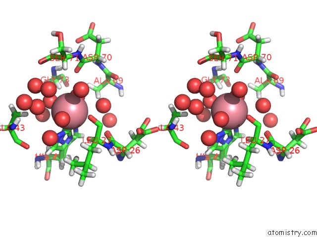
Stereo pair view
Mono view
Stereo pair view
A full contact list of Cobalt with other atoms in the Co binding
site number 1 of Crystal Structure of the E. Coli Type 1 Pilus Subunit Fimg (Engineered Variant with Substitution Q134E; N-Terminal Fimg Residues 1-12 Truncated) in Complex with the Donor Strand Peptide DSF_SRIRIRGYVR within 5.0Å range:
|
Cobalt binding site 2 out of 2 in 5iqn
Go back to
Cobalt binding site 2 out
of 2 in the Crystal Structure of the E. Coli Type 1 Pilus Subunit Fimg (Engineered Variant with Substitution Q134E; N-Terminal Fimg Residues 1-12 Truncated) in Complex with the Donor Strand Peptide DSF_SRIRIRGYVR
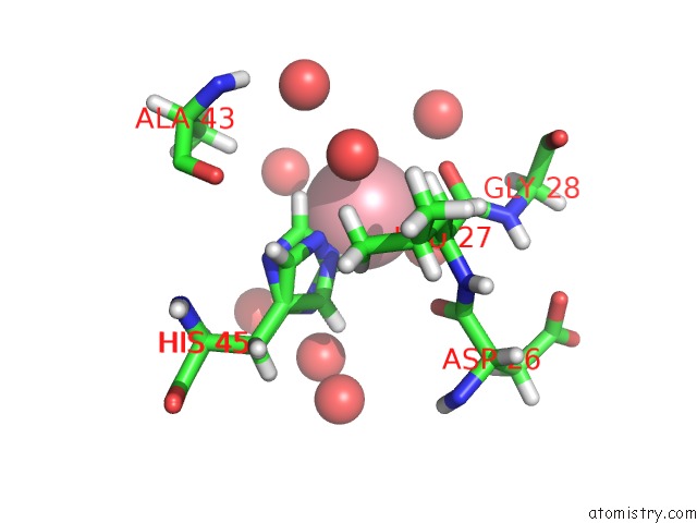
Mono view
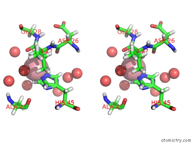
Stereo pair view
Mono view
Stereo pair view
A full contact list of Cobalt with other atoms in the Co binding
site number 2 of Crystal Structure of the E. Coli Type 1 Pilus Subunit Fimg (Engineered Variant with Substitution Q134E; N-Terminal Fimg Residues 1-12 Truncated) in Complex with the Donor Strand Peptide DSF_SRIRIRGYVR within 5.0Å range:
|
Reference:
C.Giese,
J.Eras,
A.Kern,
M.A.Scharer,
G.Capitani,
R.Glockshuber.
Accelerating the Association of the Most Stable Protein-Ligand Complex By More Than Two Orders of Magnitude. Angew.Chem.Int.Ed.Engl. V. 55 9350 2016.
ISSN: ESSN 1521-3773
PubMed: 27351462
DOI: 10.1002/ANIE.201603652
Page generated: Sun Jul 13 20:28:27 2025
ISSN: ESSN 1521-3773
PubMed: 27351462
DOI: 10.1002/ANIE.201603652
Last articles
Mg in 6KI8Mg in 6KJ6
Mg in 6KF9
Mg in 6KE2
Mg in 6KF4
Mg in 6KF3
Mg in 6KE4
Mg in 6KE0
Mg in 6KDZ
Mg in 6KDX