Cobalt »
PDB 5vtd-5zt2 »
5wro »
Cobalt in PDB 5wro: Crystal Structure of Drosophila Enolase
Enzymatic activity of Crystal Structure of Drosophila Enolase
All present enzymatic activity of Crystal Structure of Drosophila Enolase:
4.2.1.11;
4.2.1.11;
Protein crystallography data
The structure of Crystal Structure of Drosophila Enolase, PDB code: 5wro
was solved by
Z.Zhang,
Z.Shi,
with X-Ray Crystallography technique. A brief refinement statistics is given in the table below:
| Resolution Low / High (Å) | 38.33 / 2.02 |
| Space group | H 3 2 |
| Cell size a, b, c (Å), α, β, γ (°) | 118.820, 118.820, 229.792, 90.00, 90.00, 120.00 |
| R / Rfree (%) | 15.9 / 20 |
Other elements in 5wro:
The structure of Crystal Structure of Drosophila Enolase also contains other interesting chemical elements:
| Cadmium | (Cd) | 2 atoms |
| Chlorine | (Cl) | 1 atom |
Cobalt Binding Sites:
The binding sites of Cobalt atom in the Crystal Structure of Drosophila Enolase
(pdb code 5wro). This binding sites where shown within
5.0 Angstroms radius around Cobalt atom.
In total only one binding site of Cobalt was determined in the Crystal Structure of Drosophila Enolase, PDB code: 5wro:
In total only one binding site of Cobalt was determined in the Crystal Structure of Drosophila Enolase, PDB code: 5wro:
Cobalt binding site 1 out of 1 in 5wro
Go back to
Cobalt binding site 1 out
of 1 in the Crystal Structure of Drosophila Enolase
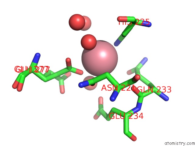
Mono view
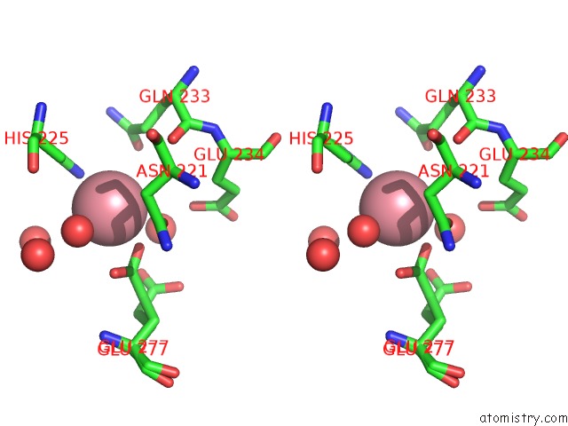
Stereo pair view
Mono view
Stereo pair view
A full contact list of Cobalt with other atoms in the Co binding
site number 1 of Crystal Structure of Drosophila Enolase within 5.0Å range:
|
Reference:
C.Sun,
B.Xu,
X.Liu,
Z.Zhang,
Z.Su.
Crystal Structure of Enolase From Drosophila Melanogaster. Acta Crystallogr F Struct V. 73 228 2017BIOL Commun.
ISSN: ESSN 2053-230X
PubMed: 28368282
DOI: 10.1107/S2053230X17004022
Page generated: Sun Jul 13 20:42:36 2025
ISSN: ESSN 2053-230X
PubMed: 28368282
DOI: 10.1107/S2053230X17004022
Last articles
Fe in 2YXOFe in 2YRS
Fe in 2YXC
Fe in 2YNM
Fe in 2YVJ
Fe in 2YP1
Fe in 2YU2
Fe in 2YU1
Fe in 2YQB
Fe in 2YOO