Cobalt »
PDB 6ega-6kfl »
6h1x »
Cobalt in PDB 6h1x: Receptor-Binding Domain of Proteus Mirabilis Uroepithelial Cell Adhesin UCAD21-211
Protein crystallography data
The structure of Receptor-Binding Domain of Proteus Mirabilis Uroepithelial Cell Adhesin UCAD21-211, PDB code: 6h1x
was solved by
J.Wangshu,
S.D.Knight,
with X-Ray Crystallography technique. A brief refinement statistics is given in the table below:
| Resolution Low / High (Å) | 41.89 / 1.70 |
| Space group | I 41 2 2 |
| Cell size a, b, c (Å), α, β, γ (°) | 73.350, 73.350, 153.106, 90.00, 90.00, 90.00 |
| R / Rfree (%) | 19.7 / 22 |
Cobalt Binding Sites:
The binding sites of Cobalt atom in the Receptor-Binding Domain of Proteus Mirabilis Uroepithelial Cell Adhesin UCAD21-211
(pdb code 6h1x). This binding sites where shown within
5.0 Angstroms radius around Cobalt atom.
In total 2 binding sites of Cobalt where determined in the Receptor-Binding Domain of Proteus Mirabilis Uroepithelial Cell Adhesin UCAD21-211, PDB code: 6h1x:
Jump to Cobalt binding site number: 1; 2;
In total 2 binding sites of Cobalt where determined in the Receptor-Binding Domain of Proteus Mirabilis Uroepithelial Cell Adhesin UCAD21-211, PDB code: 6h1x:
Jump to Cobalt binding site number: 1; 2;
Cobalt binding site 1 out of 2 in 6h1x
Go back to
Cobalt binding site 1 out
of 2 in the Receptor-Binding Domain of Proteus Mirabilis Uroepithelial Cell Adhesin UCAD21-211
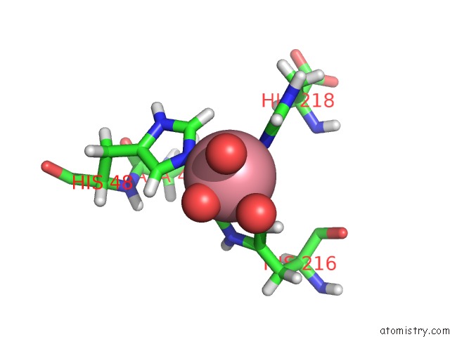
Mono view
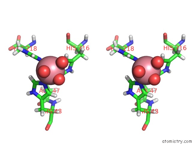
Stereo pair view
Mono view
Stereo pair view
A full contact list of Cobalt with other atoms in the Co binding
site number 1 of Receptor-Binding Domain of Proteus Mirabilis Uroepithelial Cell Adhesin UCAD21-211 within 5.0Å range:
|
Cobalt binding site 2 out of 2 in 6h1x
Go back to
Cobalt binding site 2 out
of 2 in the Receptor-Binding Domain of Proteus Mirabilis Uroepithelial Cell Adhesin UCAD21-211
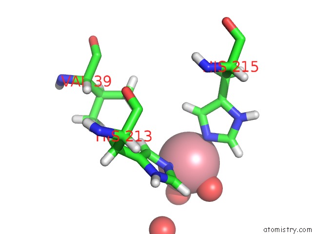
Mono view
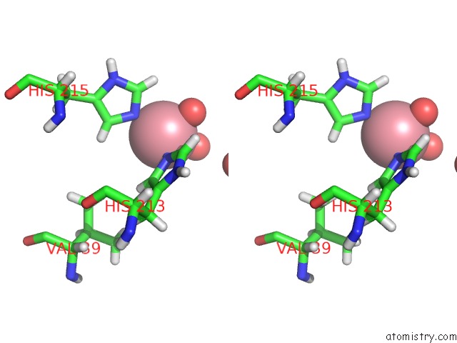
Stereo pair view
Mono view
Stereo pair view
A full contact list of Cobalt with other atoms in the Co binding
site number 2 of Receptor-Binding Domain of Proteus Mirabilis Uroepithelial Cell Adhesin UCAD21-211 within 5.0Å range:
|
Reference:
W.Jiang,
W.Ubhayasekera,
M.M.Pearson,
S.D.Knight.
Structures of Two Fimbrial Adhesins, Atfe and Ucad, From the Uropathogen Proteus Mirabilis. Acta Crystallogr D Struct V. 74 1053 2018BIOL.
ISSN: ISSN 2059-7983
PubMed: 30387764
DOI: 10.1107/S2059798318012391
Page generated: Sun Jul 13 20:57:18 2025
ISSN: ISSN 2059-7983
PubMed: 30387764
DOI: 10.1107/S2059798318012391
Last articles
Fe in 2YXOFe in 2YRS
Fe in 2YXC
Fe in 2YNM
Fe in 2YVJ
Fe in 2YP1
Fe in 2YU2
Fe in 2YU1
Fe in 2YQB
Fe in 2YOO