Cobalt »
PDB 6ega-6kfl »
6jjf »
Cobalt in PDB 6jjf: Crystal Structure of A Two-Quartet Dna Mixed-Parallel/Antiparallel G- Quadruplex
Protein crystallography data
The structure of Crystal Structure of A Two-Quartet Dna Mixed-Parallel/Antiparallel G- Quadruplex, PDB code: 6jjf
was solved by
Y.S.Zhang,
K.Ei Omari,
R.Duman,
A.Wagner,
G.N.Parkinson,
D.G.Wei,
with X-Ray Crystallography technique. A brief refinement statistics is given in the table below:
| Resolution Low / High (Å) | 23.80 / 1.47 |
| Space group | C 1 2 1 |
| Cell size a, b, c (Å), α, β, γ (°) | 45.370, 47.600, 37.730, 90.00, 110.02, 90.00 |
| R / Rfree (%) | 19.2 / 19.9 |
Other elements in 6jjf:
The structure of Crystal Structure of A Two-Quartet Dna Mixed-Parallel/Antiparallel G- Quadruplex also contains other interesting chemical elements:
| Potassium | (K) | 3 atoms |
| Sodium | (Na) | 1 atom |
Cobalt Binding Sites:
The binding sites of Cobalt atom in the Crystal Structure of A Two-Quartet Dna Mixed-Parallel/Antiparallel G- Quadruplex
(pdb code 6jjf). This binding sites where shown within
5.0 Angstroms radius around Cobalt atom.
In total 3 binding sites of Cobalt where determined in the Crystal Structure of A Two-Quartet Dna Mixed-Parallel/Antiparallel G- Quadruplex, PDB code: 6jjf:
Jump to Cobalt binding site number: 1; 2; 3;
In total 3 binding sites of Cobalt where determined in the Crystal Structure of A Two-Quartet Dna Mixed-Parallel/Antiparallel G- Quadruplex, PDB code: 6jjf:
Jump to Cobalt binding site number: 1; 2; 3;
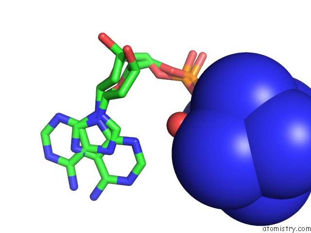
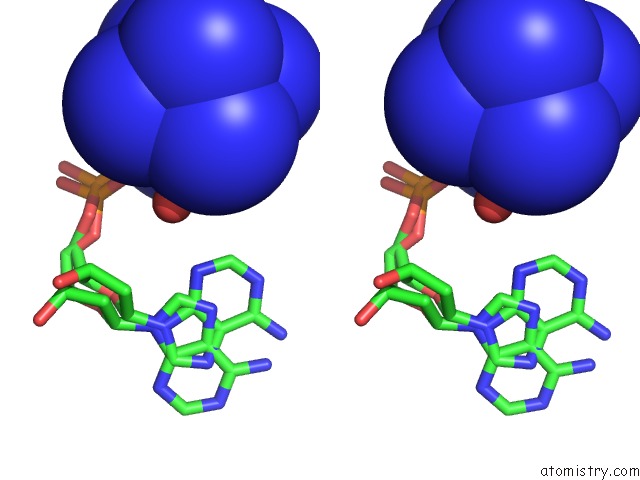
Cobalt binding site 1 out of 3 in 6jjf
Go back to
Cobalt binding site 1 out
of 3 in the Crystal Structure of A Two-Quartet Dna Mixed-Parallel/Antiparallel G- Quadruplex
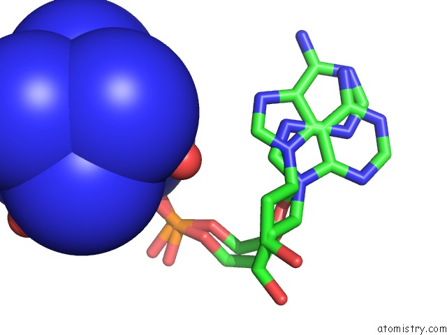
Mono view
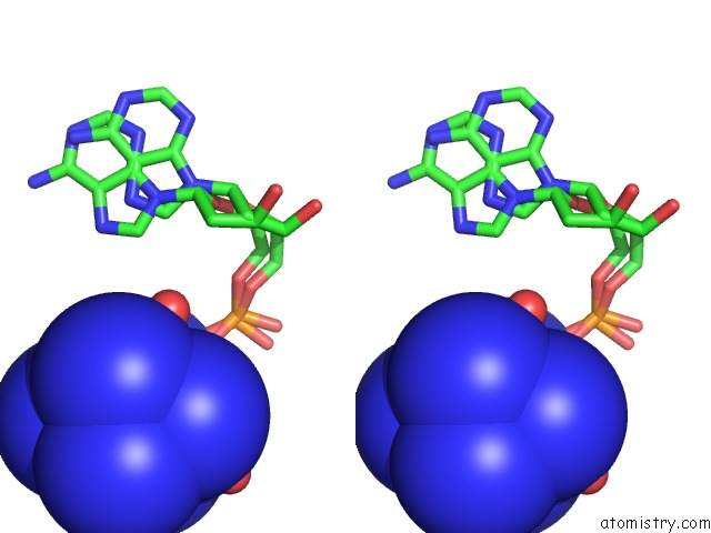
Stereo pair view
Mono view
Stereo pair view
A full contact list of Cobalt with other atoms in the Co binding
site number 1 of Crystal Structure of A Two-Quartet Dna Mixed-Parallel/Antiparallel G- Quadruplex within 5.0Å range:
|
Cobalt binding site 2 out of 3 in 6jjf
Go back to
Cobalt binding site 2 out
of 3 in the Crystal Structure of A Two-Quartet Dna Mixed-Parallel/Antiparallel G- Quadruplex
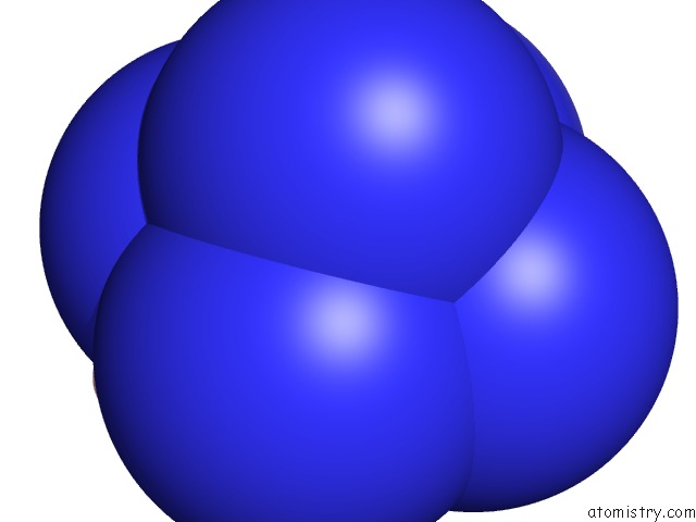
Mono view
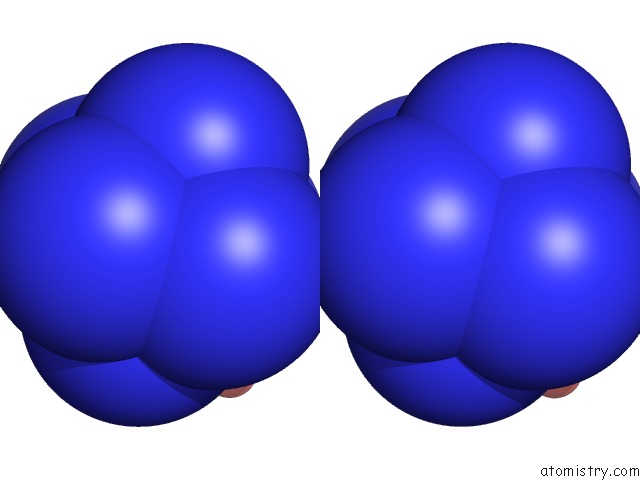
Stereo pair view
Mono view
Stereo pair view
A full contact list of Cobalt with other atoms in the Co binding
site number 2 of Crystal Structure of A Two-Quartet Dna Mixed-Parallel/Antiparallel G- Quadruplex within 5.0Å range:
|
Cobalt binding site 3 out of 3 in 6jjf
Go back to
Cobalt binding site 3 out
of 3 in the Crystal Structure of A Two-Quartet Dna Mixed-Parallel/Antiparallel G- Quadruplex
Mono view
Stereo pair view
Mono view
Stereo pair view
A full contact list of Cobalt with other atoms in the Co binding
site number 3 of Crystal Structure of A Two-Quartet Dna Mixed-Parallel/Antiparallel G- Quadruplex within 5.0Å range:
|
Reference:
Y.S.Zhang,
K.Ei Omari,
R.Duman,
A.Wagner,
G.N.Parkinson,
D.G.Wei.
Crystal Structure of A Two-Quartet Dna Mixed-Parallel/Antiparallel G-Quadruplex To Be Published.
Page generated: Sun Jul 13 21:02:07 2025
Last articles
Na in 6J0HNa in 6IWH
Na in 6IX8
Na in 6IX9
Na in 6IWG
Na in 6IX5
Na in 6IX4
Na in 6IVQ
Na in 6IVO
Na in 6IVM