Cobalt »
PDB 6ega-6kfl »
6kcx »
Cobalt in PDB 6kcx: Crystal Structure of Citrate Complex of Alpha-Glucuronidase (TM0752) From Thermotoga Maritima
Protein crystallography data
The structure of Crystal Structure of Citrate Complex of Alpha-Glucuronidase (TM0752) From Thermotoga Maritima, PDB code: 6kcx
was solved by
N.Manoj,
B.S.Mohapatra,
with X-Ray Crystallography technique. A brief refinement statistics is given in the table below:
| Resolution Low / High (Å) | 40.21 / 1.93 |
| Space group | C 1 2 1 |
| Cell size a, b, c (Å), α, β, γ (°) | 74.420, 80.410, 89.050, 90.00, 102.10, 90.00 |
| R / Rfree (%) | 14.1 / 18.4 |
Cobalt Binding Sites:
The binding sites of Cobalt atom in the Crystal Structure of Citrate Complex of Alpha-Glucuronidase (TM0752) From Thermotoga Maritima
(pdb code 6kcx). This binding sites where shown within
5.0 Angstroms radius around Cobalt atom.
In total only one binding site of Cobalt was determined in the Crystal Structure of Citrate Complex of Alpha-Glucuronidase (TM0752) From Thermotoga Maritima, PDB code: 6kcx:
In total only one binding site of Cobalt was determined in the Crystal Structure of Citrate Complex of Alpha-Glucuronidase (TM0752) From Thermotoga Maritima, PDB code: 6kcx:
Cobalt binding site 1 out of 1 in 6kcx
Go back to
Cobalt binding site 1 out
of 1 in the Crystal Structure of Citrate Complex of Alpha-Glucuronidase (TM0752) From Thermotoga Maritima
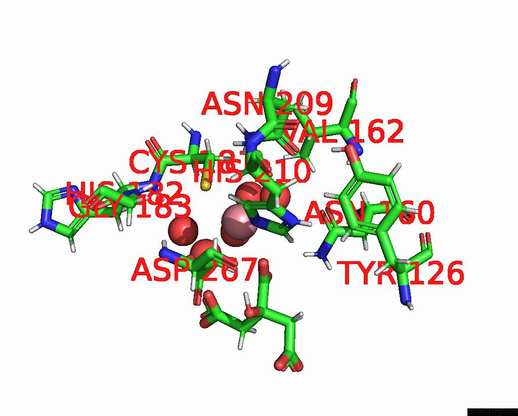
Mono view
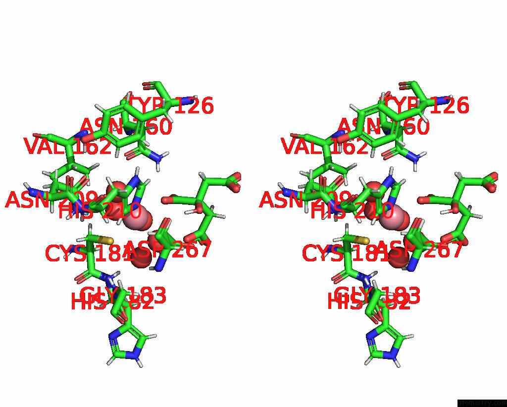
Stereo pair view
Mono view
Stereo pair view
A full contact list of Cobalt with other atoms in the Co binding
site number 1 of Crystal Structure of Citrate Complex of Alpha-Glucuronidase (TM0752) From Thermotoga Maritima within 5.0Å range:
|
Reference:
S.B.Mohapatra,
N.Manoj.
Structure of An Alpha-Glucuronidase in Complex with CO2+and Citrate Provides Insights Into the Mechanism and Substrate Recognition in the Family 4 Glycosyl Hydrolases. Biochem.Biophys.Res.Commun. V. 518 197 2019.
ISSN: ESSN 1090-2104
PubMed: 31409483
DOI: 10.1016/J.BBRC.2019.08.030
Page generated: Sun Jul 13 21:05:22 2025
ISSN: ESSN 1090-2104
PubMed: 31409483
DOI: 10.1016/J.BBRC.2019.08.030
Last articles
Fe in 2YXOFe in 2YRS
Fe in 2YXC
Fe in 2YNM
Fe in 2YVJ
Fe in 2YP1
Fe in 2YU2
Fe in 2YU1
Fe in 2YQB
Fe in 2YOO