Cobalt »
PDB 6kgh-6oxc »
6m00 »
Cobalt in PDB 6m00: Crystal Structure of Methionine Aminopeptidase From Pyrococcus Furiosus
Enzymatic activity of Crystal Structure of Methionine Aminopeptidase From Pyrococcus Furiosus
All present enzymatic activity of Crystal Structure of Methionine Aminopeptidase From Pyrococcus Furiosus:
3.4.11.18;
3.4.11.18;
Protein crystallography data
The structure of Crystal Structure of Methionine Aminopeptidase From Pyrococcus Furiosus, PDB code: 6m00
was solved by
C.B.Sandeep,
A.Addlagatta,
with X-Ray Crystallography technique. A brief refinement statistics is given in the table below:
| Resolution Low / High (Å) | 45.79 / 3.20 |
| Space group | P 62 |
| Cell size a, b, c (Å), α, β, γ (°) | 137.684, 137.684, 61.332, 90, 90, 120 |
| R / Rfree (%) | 24.3 / 27.6 |
Cobalt Binding Sites:
The binding sites of Cobalt atom in the Crystal Structure of Methionine Aminopeptidase From Pyrococcus Furiosus
(pdb code 6m00). This binding sites where shown within
5.0 Angstroms radius around Cobalt atom.
In total 2 binding sites of Cobalt where determined in the Crystal Structure of Methionine Aminopeptidase From Pyrococcus Furiosus, PDB code: 6m00:
Jump to Cobalt binding site number: 1; 2;
In total 2 binding sites of Cobalt where determined in the Crystal Structure of Methionine Aminopeptidase From Pyrococcus Furiosus, PDB code: 6m00:
Jump to Cobalt binding site number: 1; 2;
Cobalt binding site 1 out of 2 in 6m00
Go back to
Cobalt binding site 1 out
of 2 in the Crystal Structure of Methionine Aminopeptidase From Pyrococcus Furiosus
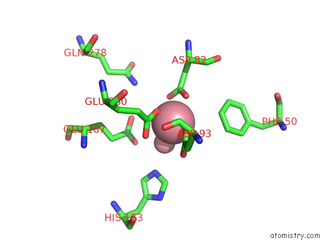
Mono view
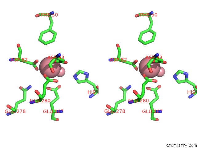
Stereo pair view
Mono view
Stereo pair view
A full contact list of Cobalt with other atoms in the Co binding
site number 1 of Crystal Structure of Methionine Aminopeptidase From Pyrococcus Furiosus within 5.0Å range:
|
Cobalt binding site 2 out of 2 in 6m00
Go back to
Cobalt binding site 2 out
of 2 in the Crystal Structure of Methionine Aminopeptidase From Pyrococcus Furiosus
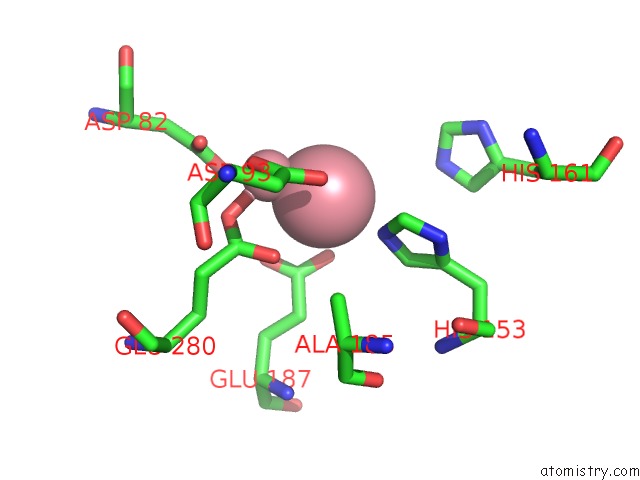
Mono view
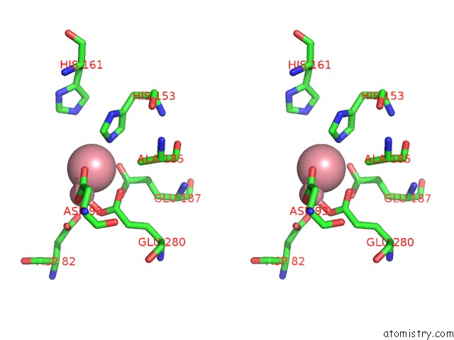
Stereo pair view
Mono view
Stereo pair view
A full contact list of Cobalt with other atoms in the Co binding
site number 2 of Crystal Structure of Methionine Aminopeptidase From Pyrococcus Furiosus within 5.0Å range:
|
Reference:
C.B.Sandeep,
A.Addlagatta.
Crystal Structure of Methionine Aminopeptidase From Pyrococcus Furiosus To Be Published.
Page generated: Sun Jul 13 21:07:33 2025
Last articles
Fe in 2YXOFe in 2YRS
Fe in 2YXC
Fe in 2YNM
Fe in 2YVJ
Fe in 2YP1
Fe in 2YU2
Fe in 2YU1
Fe in 2YQB
Fe in 2YOO