Cobalt »
PDB 7z0v-8dyl »
7zd2 »
Cobalt in PDB 7zd2: Crystal Structure of Pseudomonas Aeruginosa S-Adenosyl-L-Homocysteine Hydrolase Inhibited By CO2+ Ions.
Enzymatic activity of Crystal Structure of Pseudomonas Aeruginosa S-Adenosyl-L-Homocysteine Hydrolase Inhibited By CO2+ Ions.
All present enzymatic activity of Crystal Structure of Pseudomonas Aeruginosa S-Adenosyl-L-Homocysteine Hydrolase Inhibited By CO2+ Ions.:
3.3.1.1;
3.3.1.1;
Protein crystallography data
The structure of Crystal Structure of Pseudomonas Aeruginosa S-Adenosyl-L-Homocysteine Hydrolase Inhibited By CO2+ Ions., PDB code: 7zd2
was solved by
P.H.Malecki,
M.Gawel,
K.Brzezinski,
with X-Ray Crystallography technique. A brief refinement statistics is given in the table below:
| Resolution Low / High (Å) | 29.32 / 2.16 |
| Space group | C 1 2 1 |
| Cell size a, b, c (Å), α, β, γ (°) | 177.15, 134.21, 109.61, 90, 106.08, 90 |
| R / Rfree (%) | 18 / 22.7 |
Other elements in 7zd2:
The structure of Crystal Structure of Pseudomonas Aeruginosa S-Adenosyl-L-Homocysteine Hydrolase Inhibited By CO2+ Ions. also contains other interesting chemical elements:
| Potassium | (K) | 4 atoms |
| Chlorine | (Cl) | 1 atom |
Cobalt Binding Sites:
Pages:
>>> Page 1 <<< Page 2, Binding sites: 11 - 15;Binding sites:
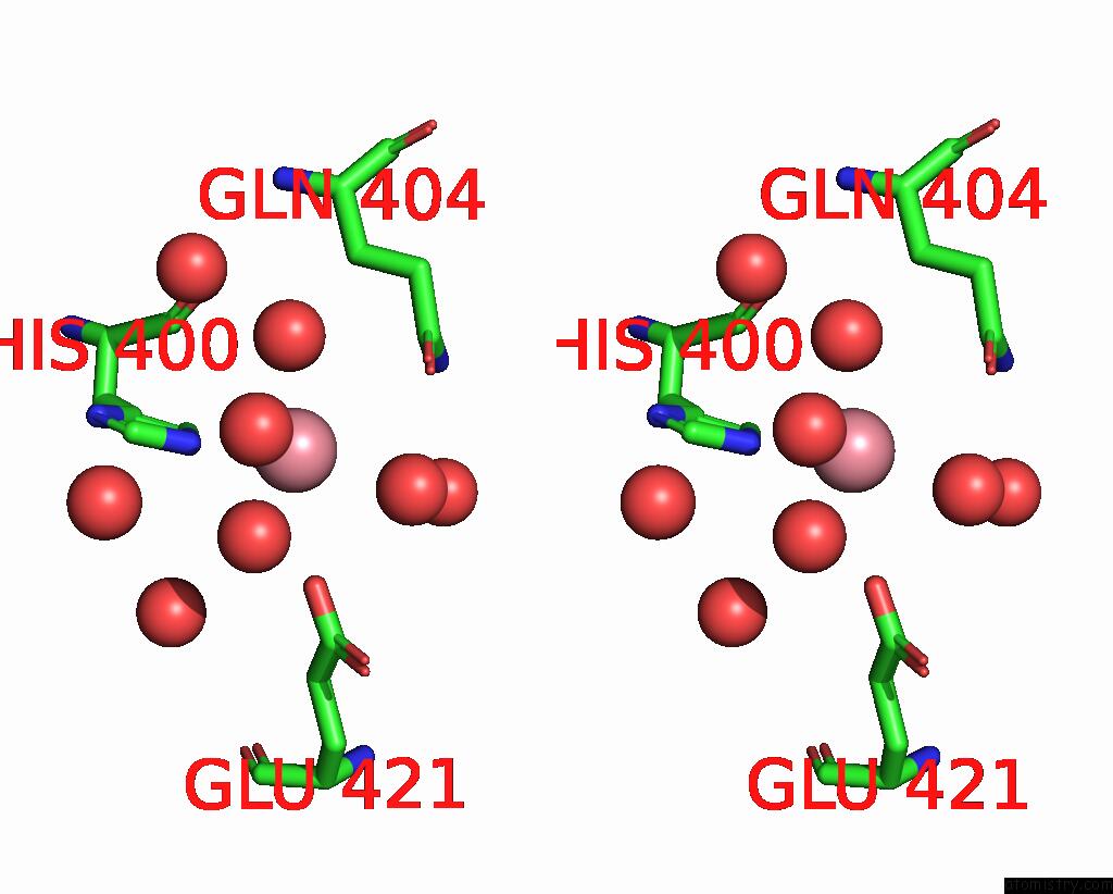
The binding sites of Cobalt atom in the Crystal Structure of Pseudomonas Aeruginosa S-Adenosyl-L-Homocysteine Hydrolase Inhibited By CO2+ Ions. (pdb code 7zd2). This binding sites where shown within 5.0 Angstroms radius around Cobalt atom.In total 15 binding sites of Cobalt where determined in the Crystal Structure of Pseudomonas Aeruginosa S-Adenosyl-L-Homocysteine Hydrolase Inhibited By CO2+ Ions., PDB code: 7zd2:
Jump to Cobalt binding site number: 1; 2; 3; 4; 5; 6; 7; 8; 9; 10;
Cobalt binding site 1 out of 15 in 7zd2
Go back to
Cobalt binding site 1 out
of 15 in the Crystal Structure of Pseudomonas Aeruginosa S-Adenosyl-L-Homocysteine Hydrolase Inhibited By CO2+ Ions.

Mono view

Stereo pair view
Mono view
Stereo pair view
A full contact list of Cobalt with other atoms in the Co binding
site number 1 of Crystal Structure of Pseudomonas Aeruginosa S-Adenosyl-L-Homocysteine Hydrolase Inhibited By CO2+ Ions. within 5.0Å range:
|
Cobalt binding site 2 out of 15 in 7zd2
Go back to
Cobalt binding site 2 out
of 15 in the Crystal Structure of Pseudomonas Aeruginosa S-Adenosyl-L-Homocysteine Hydrolase Inhibited By CO2+ Ions.

Mono view

Stereo pair view
Mono view
Stereo pair view
A full contact list of Cobalt with other atoms in the Co binding
site number 2 of Crystal Structure of Pseudomonas Aeruginosa S-Adenosyl-L-Homocysteine Hydrolase Inhibited By CO2+ Ions. within 5.0Å range:
|
Cobalt binding site 3 out of 15 in 7zd2
Go back to
Cobalt binding site 3 out
of 15 in the Crystal Structure of Pseudomonas Aeruginosa S-Adenosyl-L-Homocysteine Hydrolase Inhibited By CO2+ Ions.

Mono view

Stereo pair view
Mono view
Stereo pair view
A full contact list of Cobalt with other atoms in the Co binding
site number 3 of Crystal Structure of Pseudomonas Aeruginosa S-Adenosyl-L-Homocysteine Hydrolase Inhibited By CO2+ Ions. within 5.0Å range:
|
Cobalt binding site 4 out of 15 in 7zd2
Go back to
Cobalt binding site 4 out
of 15 in the Crystal Structure of Pseudomonas Aeruginosa S-Adenosyl-L-Homocysteine Hydrolase Inhibited By CO2+ Ions.

Mono view

Stereo pair view
Mono view
Stereo pair view
A full contact list of Cobalt with other atoms in the Co binding
site number 4 of Crystal Structure of Pseudomonas Aeruginosa S-Adenosyl-L-Homocysteine Hydrolase Inhibited By CO2+ Ions. within 5.0Å range:
|
Cobalt binding site 5 out of 15 in 7zd2
Go back to
Cobalt binding site 5 out
of 15 in the Crystal Structure of Pseudomonas Aeruginosa S-Adenosyl-L-Homocysteine Hydrolase Inhibited By CO2+ Ions.

Mono view

Stereo pair view
Mono view
Stereo pair view
A full contact list of Cobalt with other atoms in the Co binding
site number 5 of Crystal Structure of Pseudomonas Aeruginosa S-Adenosyl-L-Homocysteine Hydrolase Inhibited By CO2+ Ions. within 5.0Å range:
|
Cobalt binding site 6 out of 15 in 7zd2
Go back to
Cobalt binding site 6 out
of 15 in the Crystal Structure of Pseudomonas Aeruginosa S-Adenosyl-L-Homocysteine Hydrolase Inhibited By CO2+ Ions.

Mono view
Stereo pair view
Mono view
Stereo pair view
A full contact list of Cobalt with other atoms in the Co binding
site number 6 of Crystal Structure of Pseudomonas Aeruginosa S-Adenosyl-L-Homocysteine Hydrolase Inhibited By CO2+ Ions. within 5.0Å range:
|
Cobalt binding site 7 out of 15 in 7zd2
Go back to
Cobalt binding site 7 out
of 15 in the Crystal Structure of Pseudomonas Aeruginosa S-Adenosyl-L-Homocysteine Hydrolase Inhibited By CO2+ Ions.

Mono view

Stereo pair view
Mono view
Stereo pair view
A full contact list of Cobalt with other atoms in the Co binding
site number 7 of Crystal Structure of Pseudomonas Aeruginosa S-Adenosyl-L-Homocysteine Hydrolase Inhibited By CO2+ Ions. within 5.0Å range:
|
Cobalt binding site 8 out of 15 in 7zd2
Go back to
Cobalt binding site 8 out
of 15 in the Crystal Structure of Pseudomonas Aeruginosa S-Adenosyl-L-Homocysteine Hydrolase Inhibited By CO2+ Ions.

Mono view

Stereo pair view
Mono view
Stereo pair view
A full contact list of Cobalt with other atoms in the Co binding
site number 8 of Crystal Structure of Pseudomonas Aeruginosa S-Adenosyl-L-Homocysteine Hydrolase Inhibited By CO2+ Ions. within 5.0Å range:
|
Cobalt binding site 9 out of 15 in 7zd2
Go back to
Cobalt binding site 9 out
of 15 in the Crystal Structure of Pseudomonas Aeruginosa S-Adenosyl-L-Homocysteine Hydrolase Inhibited By CO2+ Ions.

Mono view

Stereo pair view
Mono view
Stereo pair view
A full contact list of Cobalt with other atoms in the Co binding
site number 9 of Crystal Structure of Pseudomonas Aeruginosa S-Adenosyl-L-Homocysteine Hydrolase Inhibited By CO2+ Ions. within 5.0Å range:
|
Cobalt binding site 10 out of 15 in 7zd2
Go back to
Cobalt binding site 10 out
of 15 in the Crystal Structure of Pseudomonas Aeruginosa S-Adenosyl-L-Homocysteine Hydrolase Inhibited By CO2+ Ions.

Mono view

Stereo pair view
Mono view
Stereo pair view
A full contact list of Cobalt with other atoms in the Co binding
site number 10 of Crystal Structure of Pseudomonas Aeruginosa S-Adenosyl-L-Homocysteine Hydrolase Inhibited By CO2+ Ions. within 5.0Å range:
|
Reference:
P.H.Malecki,
M.Gawel,
K.Brzezinski.
Crystal Structure of Pseudomonas Aeruginosa S-Adenosyl-L-Homocysteine Hydrolase Inhibited By CO2+ Ions. To Be Published.
Page generated: Sun Jul 13 21:47:21 2025
Last articles
Mg in 4YN0Mg in 4YMU
Mg in 4YMN
Mg in 4YLP
Mg in 4YMG
Mg in 4YLO
Mg in 4YLN
Mg in 4YLG
Mg in 4YKH
Mg in 4YKP