Cobalt »
PDB 7z0v-8dyl »
8dyl »
Cobalt in PDB 8dyl: Crystal Structure of Human Methylmalonyl-Coa Mutase Bound to Aquocobalamin
Enzymatic activity of Crystal Structure of Human Methylmalonyl-Coa Mutase Bound to Aquocobalamin
All present enzymatic activity of Crystal Structure of Human Methylmalonyl-Coa Mutase Bound to Aquocobalamin:
5.4.99.2;
5.4.99.2;
Protein crystallography data
The structure of Crystal Structure of Human Methylmalonyl-Coa Mutase Bound to Aquocobalamin, PDB code: 8dyl
was solved by
R.N.Mascarenhas,
H.Gouda,
R.Banerjee,
with X-Ray Crystallography technique. A brief refinement statistics is given in the table below:
| Resolution Low / High (Å) | 39.76 / 1.90 |
| Space group | C 2 2 21 |
| Cell size a, b, c (Å), α, β, γ (°) | 59.377, 136.07, 196.015, 90, 90, 90 |
| R / Rfree (%) | 17.5 / 20.9 |
Cobalt Binding Sites:
The binding sites of Cobalt atom in the Crystal Structure of Human Methylmalonyl-Coa Mutase Bound to Aquocobalamin
(pdb code 8dyl). This binding sites where shown within
5.0 Angstroms radius around Cobalt atom.
In total only one binding site of Cobalt was determined in the Crystal Structure of Human Methylmalonyl-Coa Mutase Bound to Aquocobalamin, PDB code: 8dyl:
In total only one binding site of Cobalt was determined in the Crystal Structure of Human Methylmalonyl-Coa Mutase Bound to Aquocobalamin, PDB code: 8dyl:
Cobalt binding site 1 out of 1 in 8dyl
Go back to
Cobalt binding site 1 out
of 1 in the Crystal Structure of Human Methylmalonyl-Coa Mutase Bound to Aquocobalamin
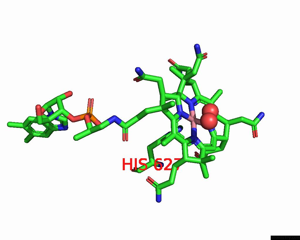
Mono view

Stereo pair view
Mono view
Stereo pair view
A full contact list of Cobalt with other atoms in the Co binding
site number 1 of Crystal Structure of Human Methylmalonyl-Coa Mutase Bound to Aquocobalamin within 5.0Å range:
|
Reference:
H.Gouda,
R.Mascarenhas,
M.Ruetz,
M.Yaw,
R.Banerjee.
Allosteric Control of Metal Redox State Regulates Coenzyme B12 Repair To Be Published.
Page generated: Sun Jul 13 21:55:24 2025
Last articles
Mg in 9ERRMg in 9ERV
Mg in 9ERS
Mg in 9ERP
Mg in 9ERC
Mg in 9ERF
Mg in 9ERE
Mg in 9ERB
Mg in 9ERD
Mg in 9ERA