Cobalt »
PDB 9fw4-9onn »
9fxj »
Cobalt in PDB 9fxj: Crystal Structure of Cobalt(II)-Substituted Double Mutant Y115E Y117E Human Glutaminyl Cyclase in Complex with Pbd-150
Enzymatic activity of Crystal Structure of Cobalt(II)-Substituted Double Mutant Y115E Y117E Human Glutaminyl Cyclase in Complex with Pbd-150
All present enzymatic activity of Crystal Structure of Cobalt(II)-Substituted Double Mutant Y115E Y117E Human Glutaminyl Cyclase in Complex with Pbd-150:
2.3.2.5;
2.3.2.5;
Protein crystallography data
The structure of Crystal Structure of Cobalt(II)-Substituted Double Mutant Y115E Y117E Human Glutaminyl Cyclase in Complex with Pbd-150, PDB code: 9fxj
was solved by
G.Tassone,
C.Pozzi,
S.Mangani,
with X-Ray Crystallography technique. A brief refinement statistics is given in the table below:
| Resolution Low / High (Å) | 74.81 / 3.06 |
| Space group | C 1 2 1 |
| Cell size a, b, c (Å), α, β, γ (°) | 86.393, 149.627, 96.158, 90, 96.82, 90 |
| R / Rfree (%) | 17.1 / 20.2 |
Cobalt Binding Sites:
The binding sites of Cobalt atom in the Crystal Structure of Cobalt(II)-Substituted Double Mutant Y115E Y117E Human Glutaminyl Cyclase in Complex with Pbd-150
(pdb code 9fxj). This binding sites where shown within
5.0 Angstroms radius around Cobalt atom.
In total 3 binding sites of Cobalt where determined in the Crystal Structure of Cobalt(II)-Substituted Double Mutant Y115E Y117E Human Glutaminyl Cyclase in Complex with Pbd-150, PDB code: 9fxj:
Jump to Cobalt binding site number: 1; 2; 3;
In total 3 binding sites of Cobalt where determined in the Crystal Structure of Cobalt(II)-Substituted Double Mutant Y115E Y117E Human Glutaminyl Cyclase in Complex with Pbd-150, PDB code: 9fxj:
Jump to Cobalt binding site number: 1; 2; 3;
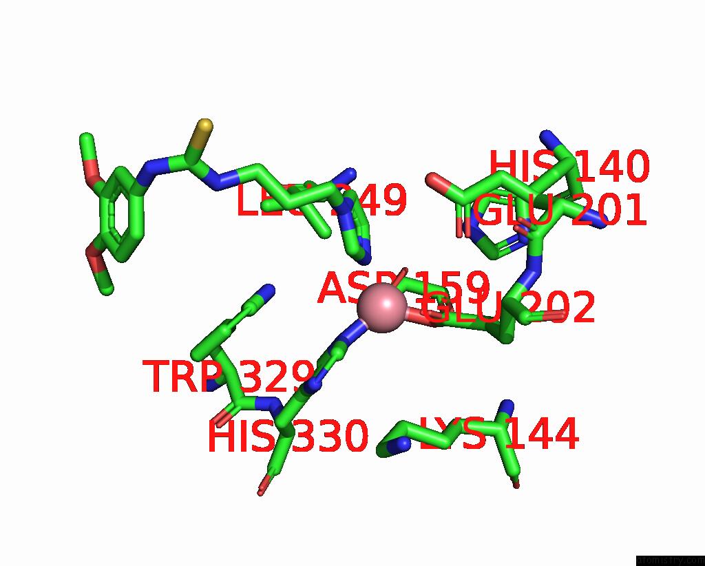
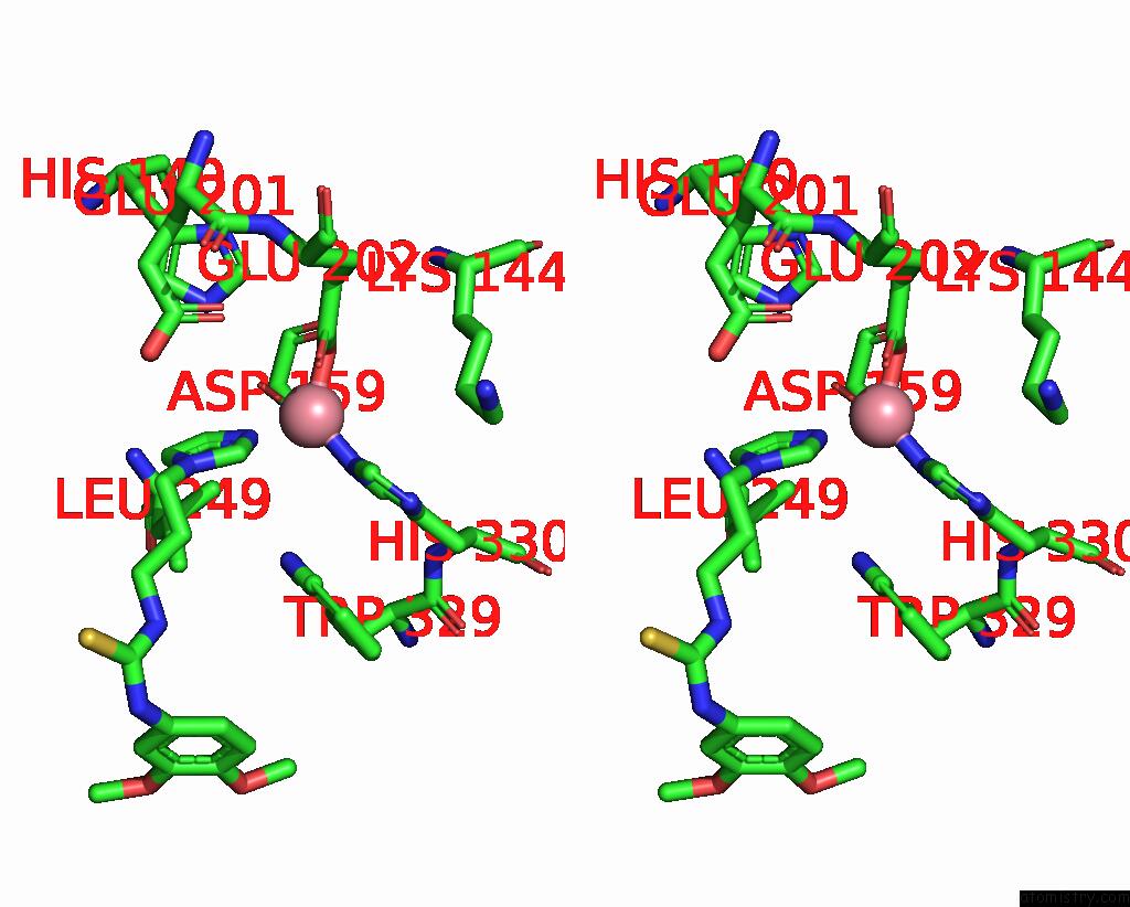
Cobalt binding site 1 out of 3 in 9fxj
Go back to
Cobalt binding site 1 out
of 3 in the Crystal Structure of Cobalt(II)-Substituted Double Mutant Y115E Y117E Human Glutaminyl Cyclase in Complex with Pbd-150
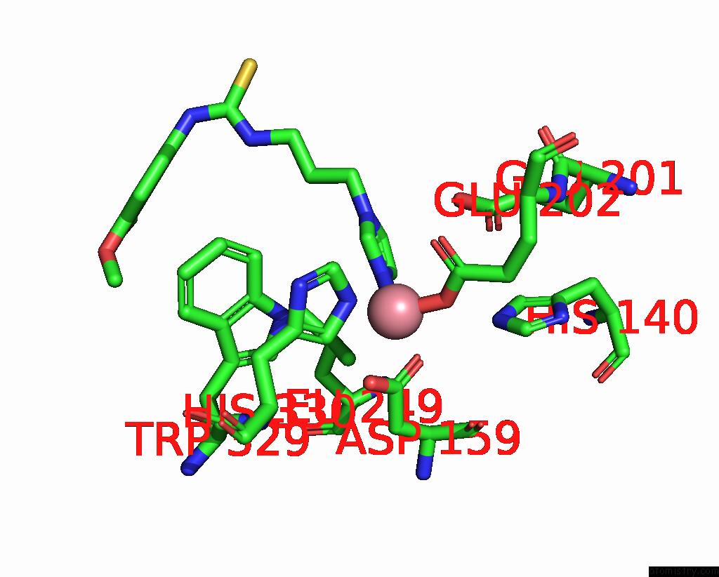
Mono view
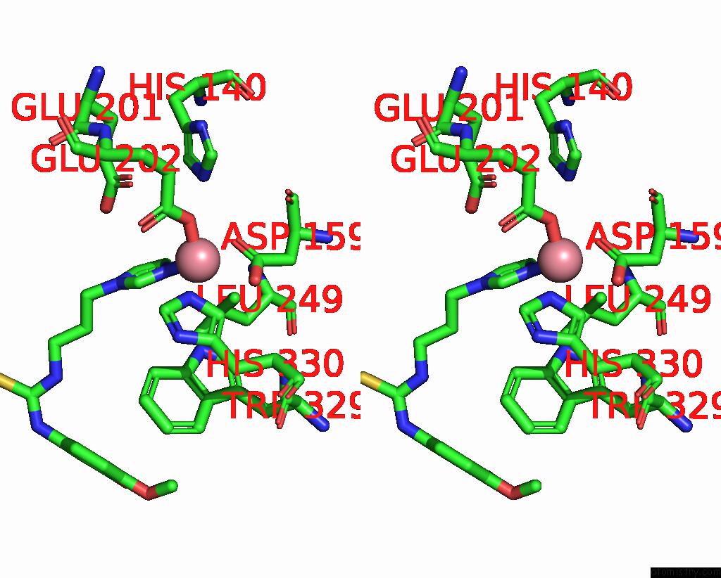
Stereo pair view
Mono view
Stereo pair view
|
|
A full contact list of Cobalt with other atoms in the Co binding
site number 1 of Crystal Structure of Cobalt(II)-Substituted Double Mutant Y115E Y117E Human Glutaminyl Cyclase in Complex with Pbd-150 within 5.0Å range:
|
Cobalt binding site 2 out of 3 in 9fxj
Go back to
Cobalt binding site 2 out
of 3 in the Crystal Structure of Cobalt(II)-Substituted Double Mutant Y115E Y117E Human Glutaminyl Cyclase in Complex with Pbd-150
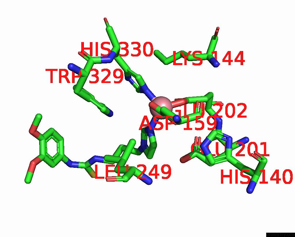
Mono view
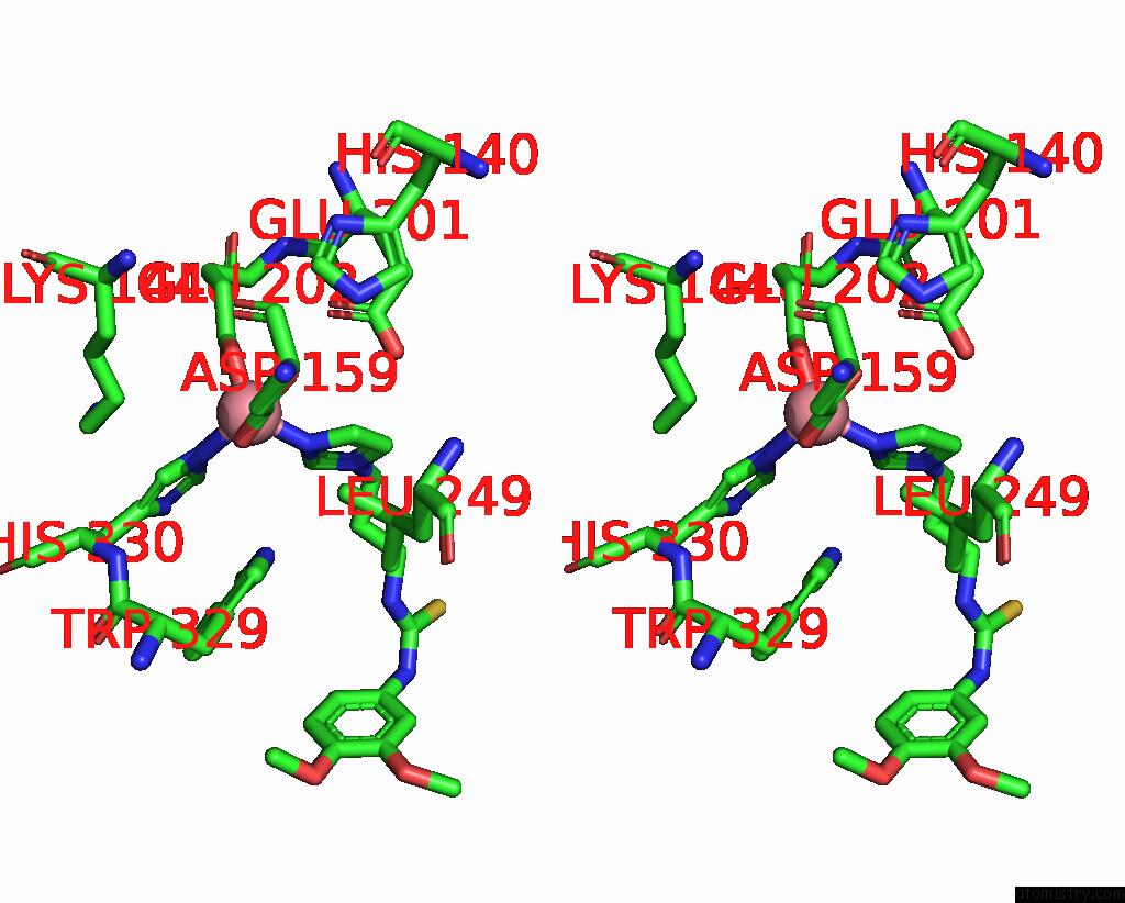
Stereo pair view
Mono view
Stereo pair view
|
|
A full contact list of Cobalt with other atoms in the Co binding
site number 2 of Crystal Structure of Cobalt(II)-Substituted Double Mutant Y115E Y117E Human Glutaminyl Cyclase in Complex with Pbd-150 within 5.0Å range:
|
Cobalt binding site 3 out of 3 in 9fxj
Go back to
Cobalt binding site 3 out
of 3 in the Crystal Structure of Cobalt(II)-Substituted Double Mutant Y115E Y117E Human Glutaminyl Cyclase in Complex with Pbd-150
Mono view
Stereo pair view
Mono view
Stereo pair view
|
|
A full contact list of Cobalt with other atoms in the Co binding
site number 3 of Crystal Structure of Cobalt(II)-Substituted Double Mutant Y115E Y117E Human Glutaminyl Cyclase in Complex with Pbd-150 within 5.0Å range:
|
Reference:
G.Tassone,
C.Pozzi,
S.Mangani.
Metal Ion Binding to Human Glutaminyl Cyclase: A Structural Perspective. Int J Mol Sci V. 25 2024.
ISSN: ESSN 1422-0067
PubMed: 39125848
DOI: 10.3390/IJMS25158279
Page generated: Sun Jul 13 22:15:02 2025
ISSN: ESSN 1422-0067
PubMed: 39125848
DOI: 10.3390/IJMS25158279
Last articles
Mg in 3VATMg in 3VA8
Mg in 3VAD
Mg in 3V9X
Mg in 3V7E
Mg in 3V9Z
Mg in 3VA0
Mg in 3V9W
Mg in 3V9U
Mg in 3V94