Cobalt »
PDB 9fvs-9onn »
9fy4 »
Cobalt in PDB 9fy4: Crystal Structure of Heme-Oxygenase Mutant I143K From Corynebacterium Diphtheriae Complexed with Cobalt-Porphyrine (Humo-Co(III))
Enzymatic activity of Crystal Structure of Heme-Oxygenase Mutant I143K From Corynebacterium Diphtheriae Complexed with Cobalt-Porphyrine (Humo-Co(III))
All present enzymatic activity of Crystal Structure of Heme-Oxygenase Mutant I143K From Corynebacterium Diphtheriae Complexed with Cobalt-Porphyrine (Humo-Co(III)):
1.14.14.18;
1.14.14.18;
Protein crystallography data
The structure of Crystal Structure of Heme-Oxygenase Mutant I143K From Corynebacterium Diphtheriae Complexed with Cobalt-Porphyrine (Humo-Co(III)), PDB code: 9fy4
was solved by
R.J.Labidi,
B.Faivre,
P.Carpentier,
J.Perard,
P.Gotico,
Y.Li,
M.Atta,
M.Fontecave,
with X-Ray Crystallography technique. A brief refinement statistics is given in the table below:
| Resolution Low / High (Å) | 48.61 / 2.84 |
| Space group | P 1 21 1 |
| Cell size a, b, c (Å), α, β, γ (°) | 41.681, 63.525, 76.12, 90, 97.37, 90 |
| R / Rfree (%) | 23.4 / 30.6 |
Other elements in 9fy4:
The structure of Crystal Structure of Heme-Oxygenase Mutant I143K From Corynebacterium Diphtheriae Complexed with Cobalt-Porphyrine (Humo-Co(III)) also contains other interesting chemical elements:
| Chlorine | (Cl) | 1 atom |
Cobalt Binding Sites:
The binding sites of Cobalt atom in the Crystal Structure of Heme-Oxygenase Mutant I143K From Corynebacterium Diphtheriae Complexed with Cobalt-Porphyrine (Humo-Co(III))
(pdb code 9fy4). This binding sites where shown within
5.0 Angstroms radius around Cobalt atom.
In total 3 binding sites of Cobalt where determined in the Crystal Structure of Heme-Oxygenase Mutant I143K From Corynebacterium Diphtheriae Complexed with Cobalt-Porphyrine (Humo-Co(III)), PDB code: 9fy4:
Jump to Cobalt binding site number: 1; 2; 3;
In total 3 binding sites of Cobalt where determined in the Crystal Structure of Heme-Oxygenase Mutant I143K From Corynebacterium Diphtheriae Complexed with Cobalt-Porphyrine (Humo-Co(III)), PDB code: 9fy4:
Jump to Cobalt binding site number: 1; 2; 3;
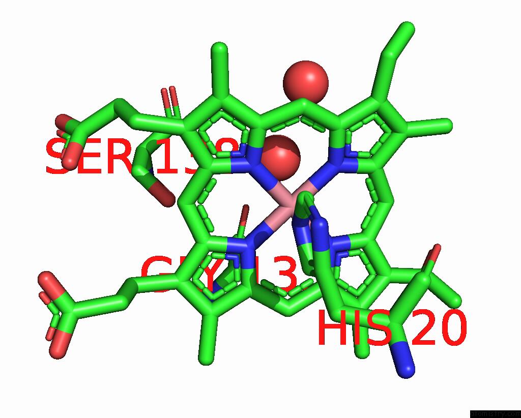
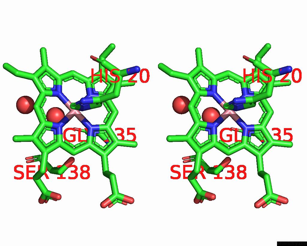
Cobalt binding site 1 out of 3 in 9fy4
Go back to
Cobalt binding site 1 out
of 3 in the Crystal Structure of Heme-Oxygenase Mutant I143K From Corynebacterium Diphtheriae Complexed with Cobalt-Porphyrine (Humo-Co(III))
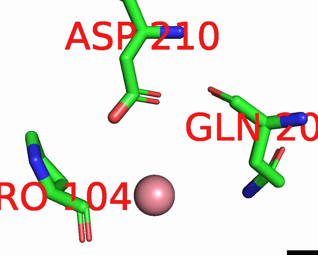
Mono view
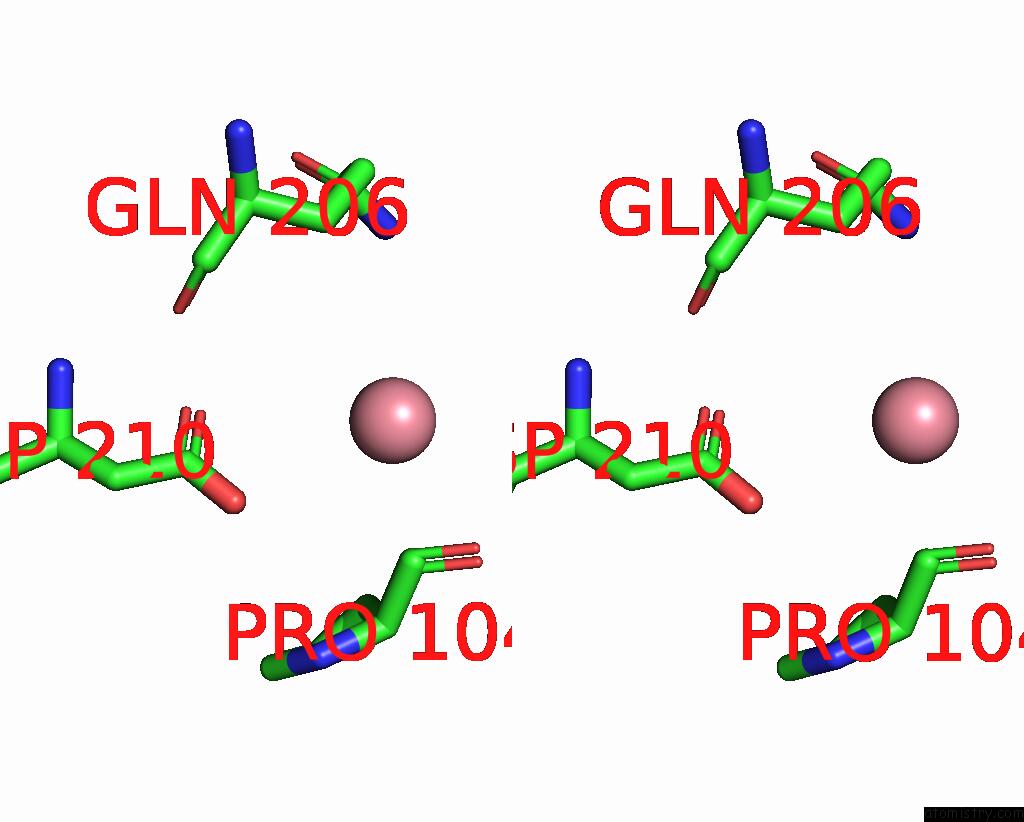
Stereo pair view
Mono view
Stereo pair view
A full contact list of Cobalt with other atoms in the Co binding
site number 1 of Crystal Structure of Heme-Oxygenase Mutant I143K From Corynebacterium Diphtheriae Complexed with Cobalt-Porphyrine (Humo-Co(III)) within 5.0Å range:
|
Cobalt binding site 2 out of 3 in 9fy4
Go back to
Cobalt binding site 2 out
of 3 in the Crystal Structure of Heme-Oxygenase Mutant I143K From Corynebacterium Diphtheriae Complexed with Cobalt-Porphyrine (Humo-Co(III))
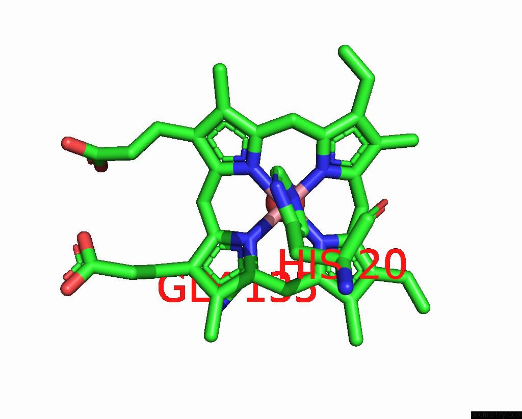
Mono view
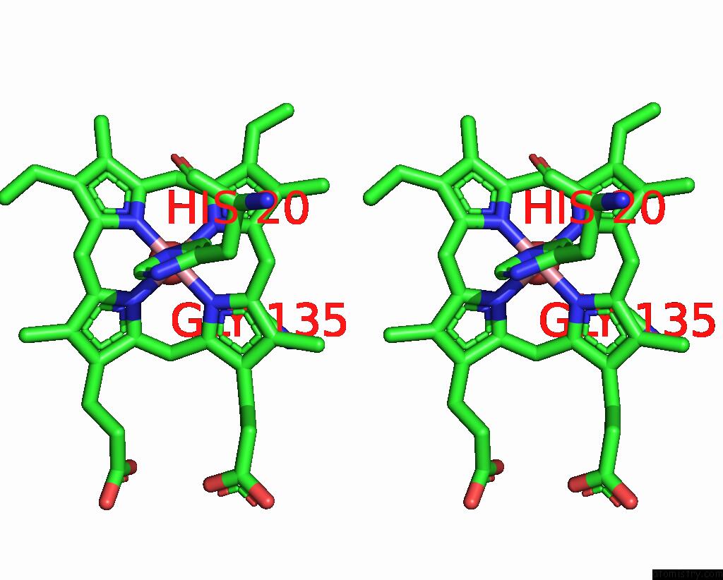
Stereo pair view
Mono view
Stereo pair view
A full contact list of Cobalt with other atoms in the Co binding
site number 2 of Crystal Structure of Heme-Oxygenase Mutant I143K From Corynebacterium Diphtheriae Complexed with Cobalt-Porphyrine (Humo-Co(III)) within 5.0Å range:
|
Cobalt binding site 3 out of 3 in 9fy4
Go back to
Cobalt binding site 3 out
of 3 in the Crystal Structure of Heme-Oxygenase Mutant I143K From Corynebacterium Diphtheriae Complexed with Cobalt-Porphyrine (Humo-Co(III))
Mono view
Stereo pair view
Mono view
Stereo pair view
A full contact list of Cobalt with other atoms in the Co binding
site number 3 of Crystal Structure of Heme-Oxygenase Mutant I143K From Corynebacterium Diphtheriae Complexed with Cobalt-Porphyrine (Humo-Co(III)) within 5.0Å range:
|
Reference:
R.J.Labidi,
B.Faivre,
P.Carpentier,
J.Perard,
P.Gotico,
Y.Li,
M.Atta,
M.Fontecave.
Light-Activated Artificial Co 2 -Reductase: Structure and Activity. J.Am.Chem.Soc. 2024.
ISSN: ESSN 1520-5126
PubMed: 39352411
DOI: 10.1021/JACS.4C08927
Page generated: Sun Jul 13 22:15:43 2025
ISSN: ESSN 1520-5126
PubMed: 39352411
DOI: 10.1021/JACS.4C08927
Last articles
Zn in 9QM9Zn in 9S44
Zn in 9OFE
Zn in 9OFC
Zn in 9OFD
Zn in 9OF1
Zn in 9OFB
Zn in 9N0J
Zn in 9M5X
Zn in 9LGI