Cobalt »
PDB 9fvs-9onn »
9grd »
Cobalt in PDB 9grd: Cryo-Electron Microscopy Structure of Glucose/Xylose Isomerase From Streptomyces Rubiginosus with Cobalt Ions in the Active Site
Enzymatic activity of Cryo-Electron Microscopy Structure of Glucose/Xylose Isomerase From Streptomyces Rubiginosus with Cobalt Ions in the Active Site
All present enzymatic activity of Cryo-Electron Microscopy Structure of Glucose/Xylose Isomerase From Streptomyces Rubiginosus with Cobalt Ions in the Active Site:
5.3.1.5;
5.3.1.5;
Cobalt Binding Sites:
The binding sites of Cobalt atom in the Cryo-Electron Microscopy Structure of Glucose/Xylose Isomerase From Streptomyces Rubiginosus with Cobalt Ions in the Active Site
(pdb code 9grd). This binding sites where shown within
5.0 Angstroms radius around Cobalt atom.
In total 8 binding sites of Cobalt where determined in the Cryo-Electron Microscopy Structure of Glucose/Xylose Isomerase From Streptomyces Rubiginosus with Cobalt Ions in the Active Site, PDB code: 9grd:
Jump to Cobalt binding site number: 1; 2; 3; 4; 5; 6; 7; 8;
In total 8 binding sites of Cobalt where determined in the Cryo-Electron Microscopy Structure of Glucose/Xylose Isomerase From Streptomyces Rubiginosus with Cobalt Ions in the Active Site, PDB code: 9grd:
Jump to Cobalt binding site number: 1; 2; 3; 4; 5; 6; 7; 8;
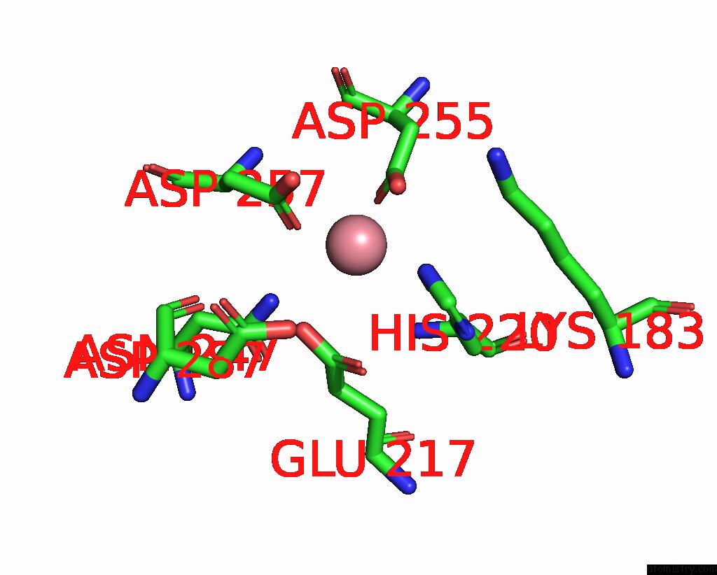
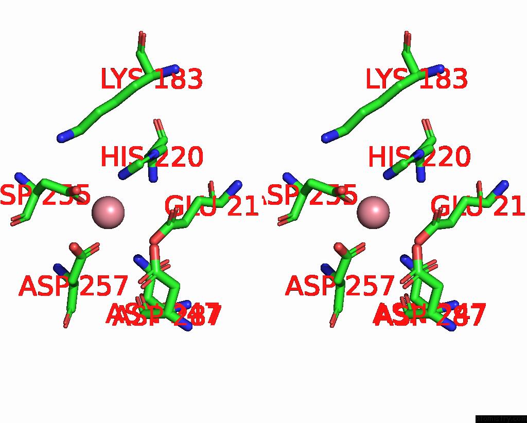
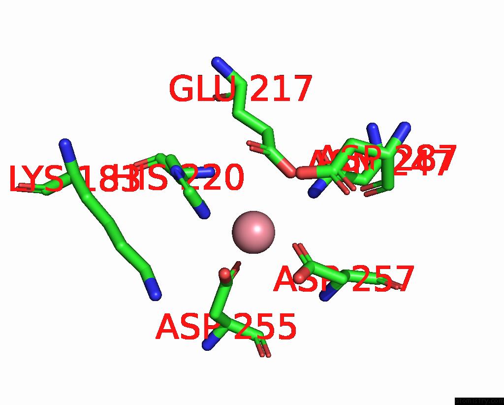
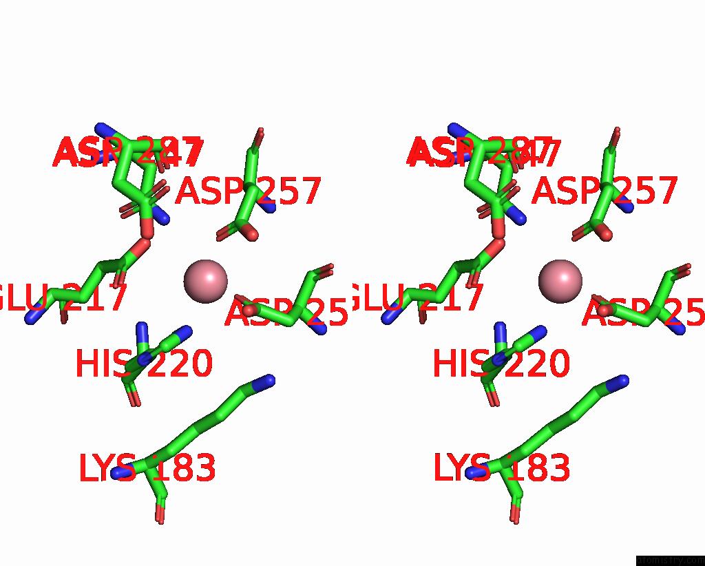
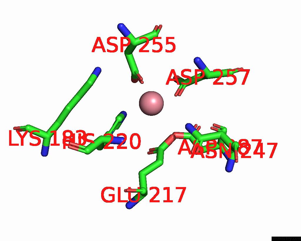
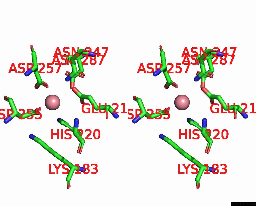
Cobalt binding site 1 out of 8 in 9grd
Go back to
Cobalt binding site 1 out
of 8 in the Cryo-Electron Microscopy Structure of Glucose/Xylose Isomerase From Streptomyces Rubiginosus with Cobalt Ions in the Active Site
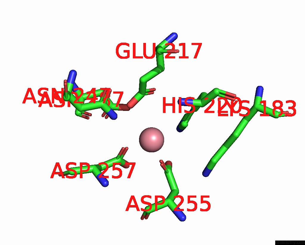
Mono view
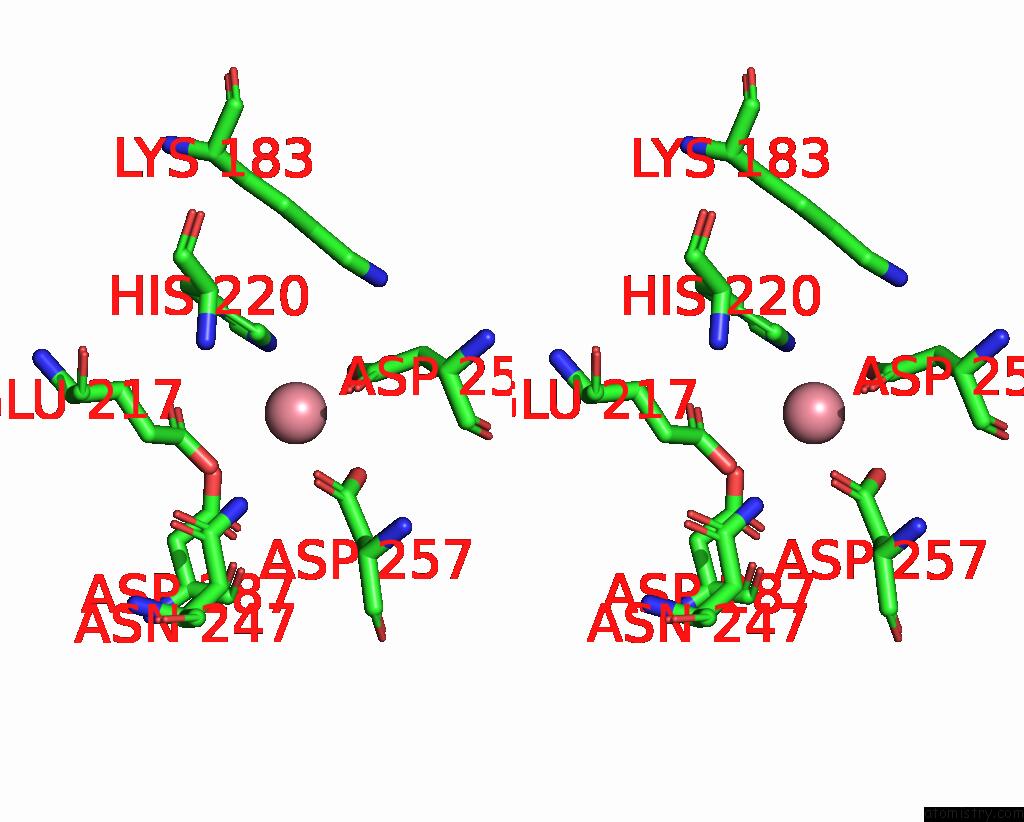
Stereo pair view
Mono view
Stereo pair view
A full contact list of Cobalt with other atoms in the Co binding
site number 1 of Cryo-Electron Microscopy Structure of Glucose/Xylose Isomerase From Streptomyces Rubiginosus with Cobalt Ions in the Active Site within 5.0Å range:
|
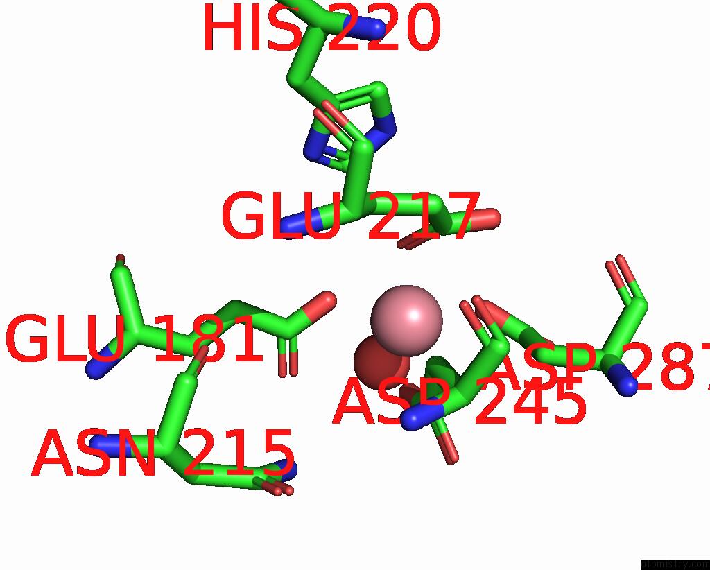
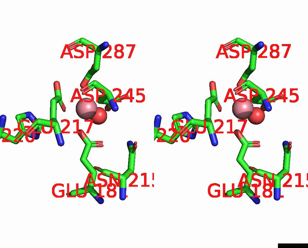
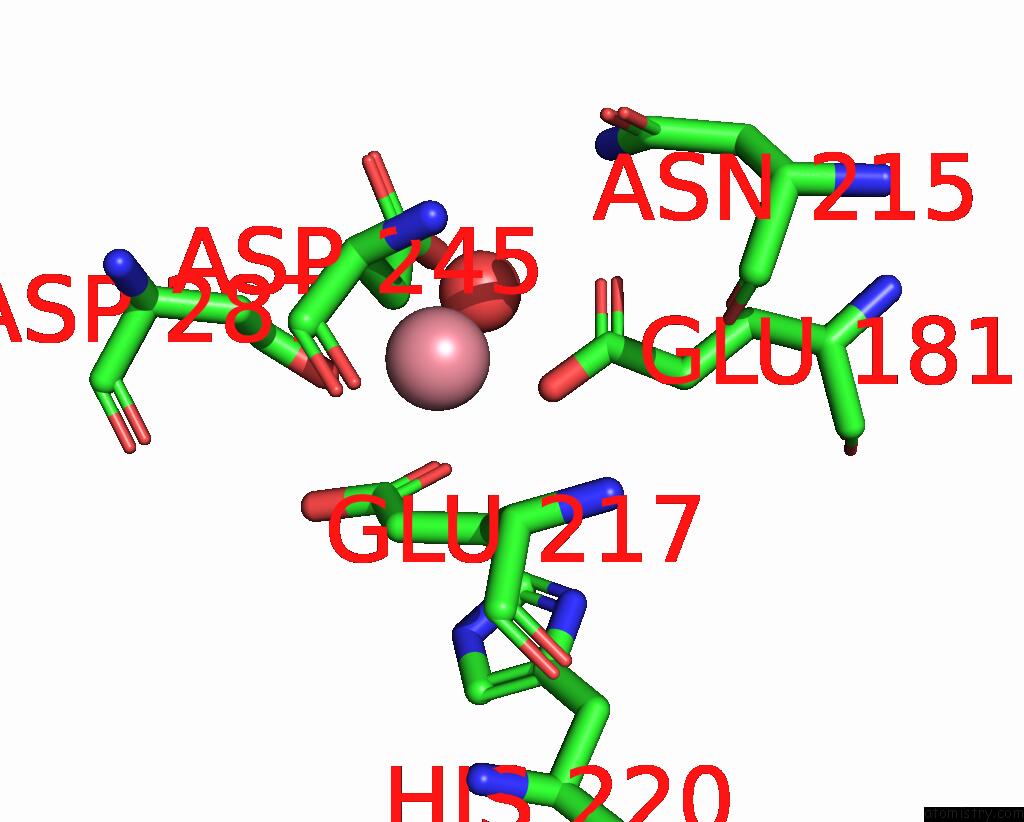
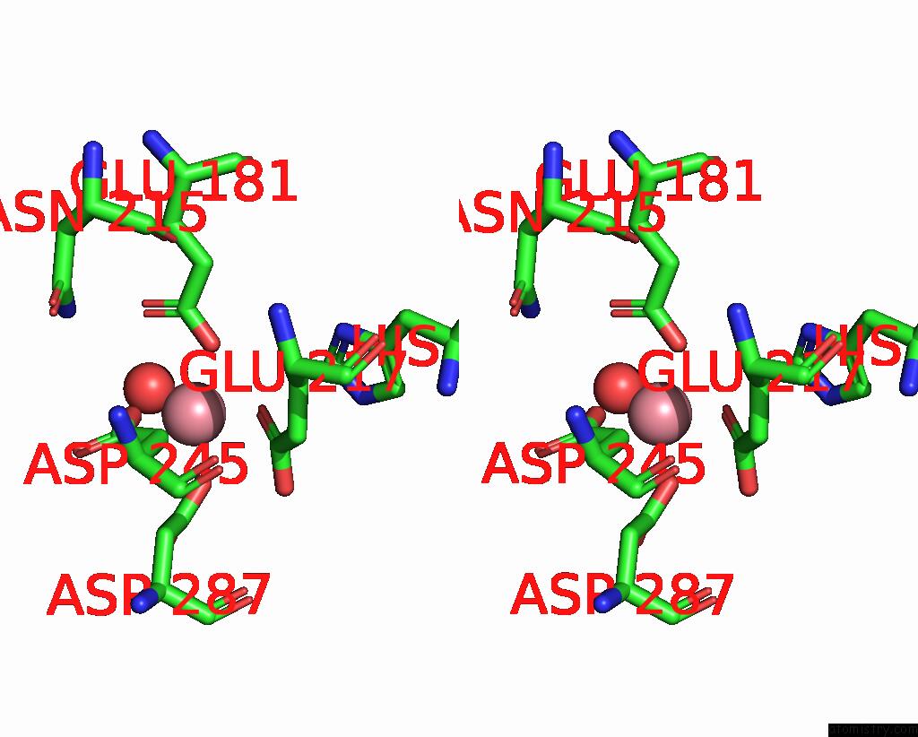
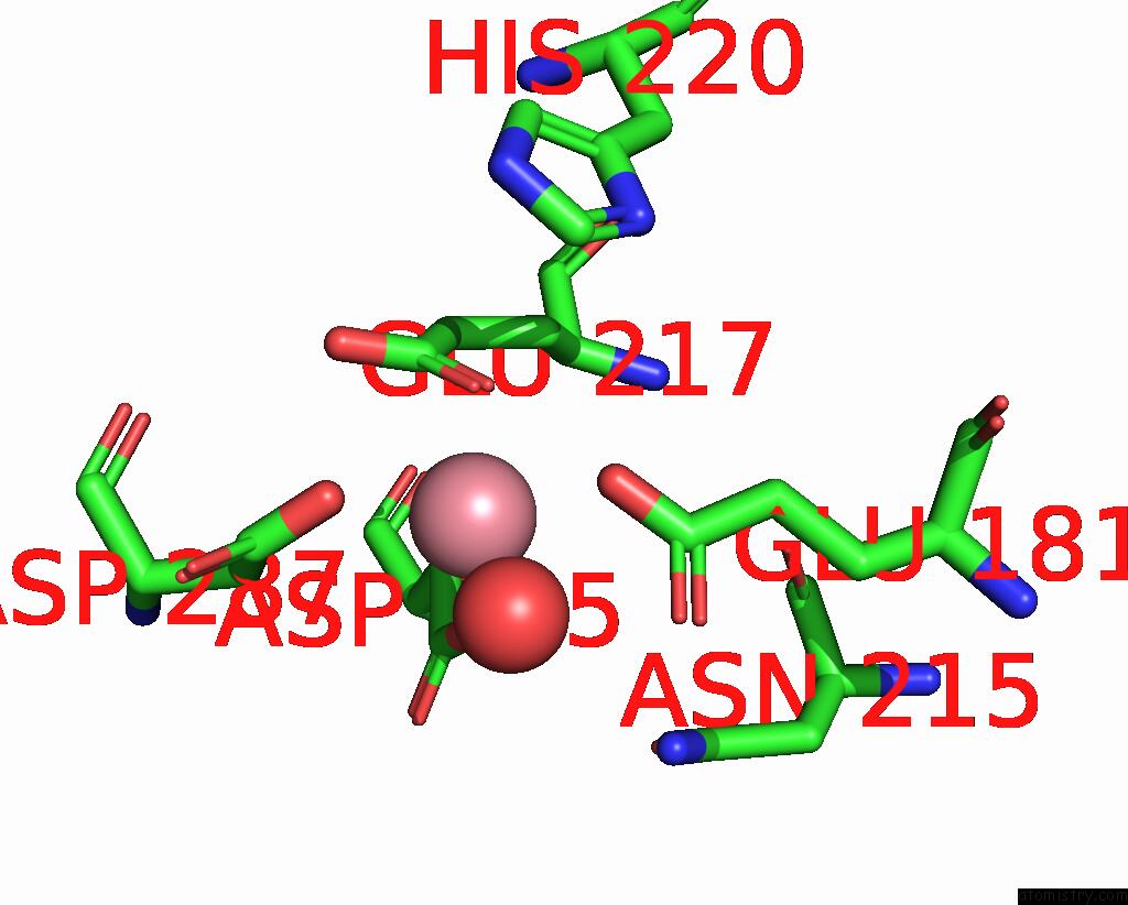
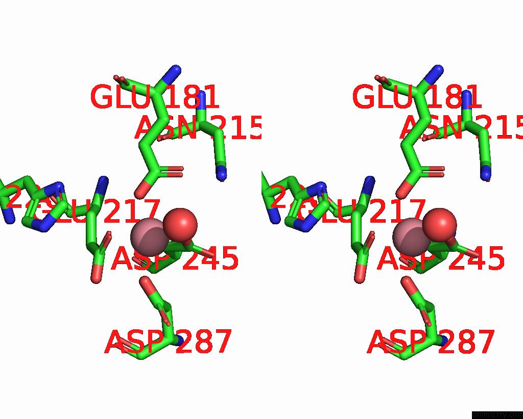
Cobalt binding site 2 out of 8 in 9grd
Go back to
Cobalt binding site 2 out
of 8 in the Cryo-Electron Microscopy Structure of Glucose/Xylose Isomerase From Streptomyces Rubiginosus with Cobalt Ions in the Active Site
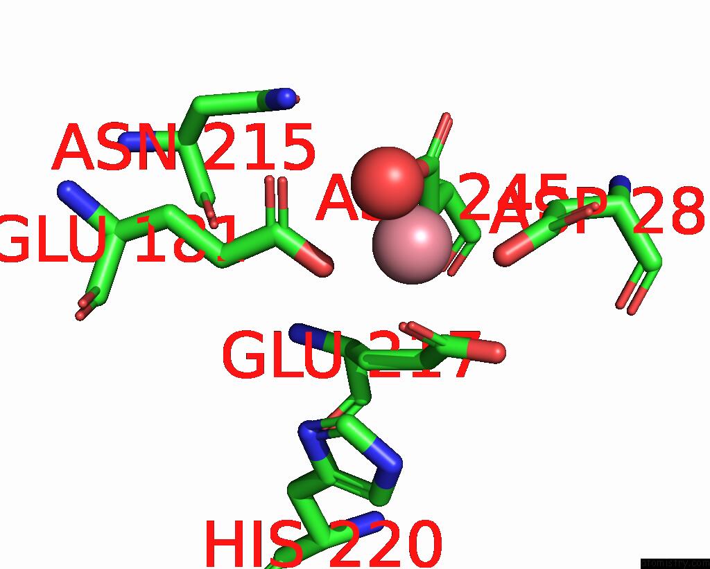
Mono view
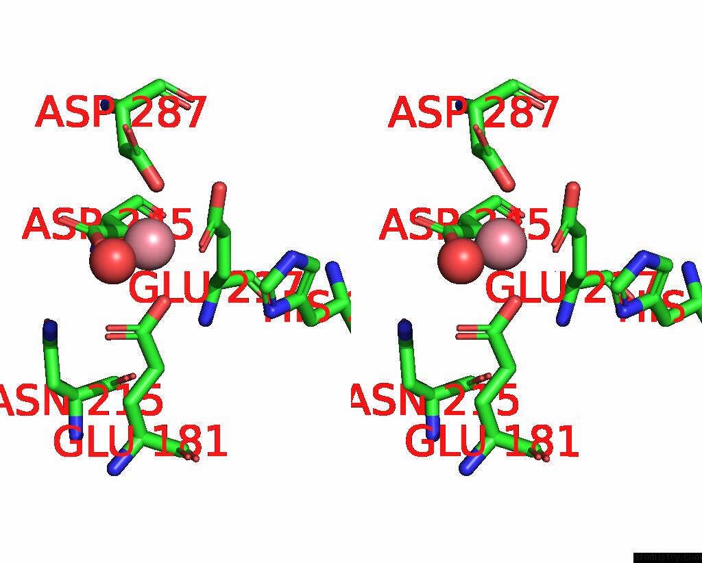
Stereo pair view
Mono view
Stereo pair view
A full contact list of Cobalt with other atoms in the Co binding
site number 2 of Cryo-Electron Microscopy Structure of Glucose/Xylose Isomerase From Streptomyces Rubiginosus with Cobalt Ions in the Active Site within 5.0Å range:
|
Cobalt binding site 3 out of 8 in 9grd
Go back to
Cobalt binding site 3 out
of 8 in the Cryo-Electron Microscopy Structure of Glucose/Xylose Isomerase From Streptomyces Rubiginosus with Cobalt Ions in the Active Site
Mono view
Stereo pair view
Mono view
Stereo pair view
A full contact list of Cobalt with other atoms in the Co binding
site number 3 of Cryo-Electron Microscopy Structure of Glucose/Xylose Isomerase From Streptomyces Rubiginosus with Cobalt Ions in the Active Site within 5.0Å range:
|
Cobalt binding site 4 out of 8 in 9grd
Go back to
Cobalt binding site 4 out
of 8 in the Cryo-Electron Microscopy Structure of Glucose/Xylose Isomerase From Streptomyces Rubiginosus with Cobalt Ions in the Active Site
Mono view
Stereo pair view
Mono view
Stereo pair view
A full contact list of Cobalt with other atoms in the Co binding
site number 4 of Cryo-Electron Microscopy Structure of Glucose/Xylose Isomerase From Streptomyces Rubiginosus with Cobalt Ions in the Active Site within 5.0Å range:
|
Cobalt binding site 5 out of 8 in 9grd
Go back to
Cobalt binding site 5 out
of 8 in the Cryo-Electron Microscopy Structure of Glucose/Xylose Isomerase From Streptomyces Rubiginosus with Cobalt Ions in the Active Site
Mono view
Stereo pair view
Mono view
Stereo pair view
A full contact list of Cobalt with other atoms in the Co binding
site number 5 of Cryo-Electron Microscopy Structure of Glucose/Xylose Isomerase From Streptomyces Rubiginosus with Cobalt Ions in the Active Site within 5.0Å range:
|
Cobalt binding site 6 out of 8 in 9grd
Go back to
Cobalt binding site 6 out
of 8 in the Cryo-Electron Microscopy Structure of Glucose/Xylose Isomerase From Streptomyces Rubiginosus with Cobalt Ions in the Active Site
Mono view
Stereo pair view
Mono view
Stereo pair view
A full contact list of Cobalt with other atoms in the Co binding
site number 6 of Cryo-Electron Microscopy Structure of Glucose/Xylose Isomerase From Streptomyces Rubiginosus with Cobalt Ions in the Active Site within 5.0Å range:
|
Cobalt binding site 7 out of 8 in 9grd
Go back to
Cobalt binding site 7 out
of 8 in the Cryo-Electron Microscopy Structure of Glucose/Xylose Isomerase From Streptomyces Rubiginosus with Cobalt Ions in the Active Site
Mono view
Stereo pair view
Mono view
Stereo pair view
A full contact list of Cobalt with other atoms in the Co binding
site number 7 of Cryo-Electron Microscopy Structure of Glucose/Xylose Isomerase From Streptomyces Rubiginosus with Cobalt Ions in the Active Site within 5.0Å range:
|
Cobalt binding site 8 out of 8 in 9grd
Go back to
Cobalt binding site 8 out
of 8 in the Cryo-Electron Microscopy Structure of Glucose/Xylose Isomerase From Streptomyces Rubiginosus with Cobalt Ions in the Active Site
Mono view
Stereo pair view
Mono view
Stereo pair view
A full contact list of Cobalt with other atoms in the Co binding
site number 8 of Cryo-Electron Microscopy Structure of Glucose/Xylose Isomerase From Streptomyces Rubiginosus with Cobalt Ions in the Active Site within 5.0Å range:
|
Reference:
J.Slawek,
A.Klonecka,
M.Kozak,
M.Rawski.
Cryo-Electron Microscopy Structure of Glucose/Xylose Isomerase From Streptomyces Rubiginosus with Cobalt Ions in the Active Site To Be Published.
Page generated: Sun Jul 13 22:16:14 2025
Last articles
Zn in 9QM9Zn in 9S44
Zn in 9OFE
Zn in 9OFC
Zn in 9OFD
Zn in 9OF1
Zn in 9OFB
Zn in 9N0J
Zn in 9M5X
Zn in 9LGI