Cobalt »
PDB 1e31-1hgw »
1egm »
Cobalt in PDB 1egm: Crystal Structure of Diol Dehydratase-Cyanocobalamin Complex at 100K.
Enzymatic activity of Crystal Structure of Diol Dehydratase-Cyanocobalamin Complex at 100K.
All present enzymatic activity of Crystal Structure of Diol Dehydratase-Cyanocobalamin Complex at 100K.:
4.2.1.28;
4.2.1.28;
Protein crystallography data
The structure of Crystal Structure of Diol Dehydratase-Cyanocobalamin Complex at 100K., PDB code: 1egm
was solved by
J.Masuda,
N.Shibata,
T.Toraya,
Y.Morimoto,
N.Yasuoka,
with X-Ray Crystallography technique. A brief refinement statistics is given in the table below:
| Resolution Low / High (Å) | 30.00 / 1.85 |
| Space group | P 21 21 21 |
| Cell size a, b, c (Å), α, β, γ (°) | 74.000, 121.600, 207.700, 90.00, 90.00, 90.00 |
| R / Rfree (%) | 18.2 / 24.9 |
Other elements in 1egm:
The structure of Crystal Structure of Diol Dehydratase-Cyanocobalamin Complex at 100K. also contains other interesting chemical elements:
| Potassium | (K) | 2 atoms |
Cobalt Binding Sites:
The binding sites of Cobalt atom in the Crystal Structure of Diol Dehydratase-Cyanocobalamin Complex at 100K.
(pdb code 1egm). This binding sites where shown within
5.0 Angstroms radius around Cobalt atom.
In total 2 binding sites of Cobalt where determined in the Crystal Structure of Diol Dehydratase-Cyanocobalamin Complex at 100K., PDB code: 1egm:
Jump to Cobalt binding site number: 1; 2;
In total 2 binding sites of Cobalt where determined in the Crystal Structure of Diol Dehydratase-Cyanocobalamin Complex at 100K., PDB code: 1egm:
Jump to Cobalt binding site number: 1; 2;
Cobalt binding site 1 out of 2 in 1egm
Go back to
Cobalt binding site 1 out
of 2 in the Crystal Structure of Diol Dehydratase-Cyanocobalamin Complex at 100K.
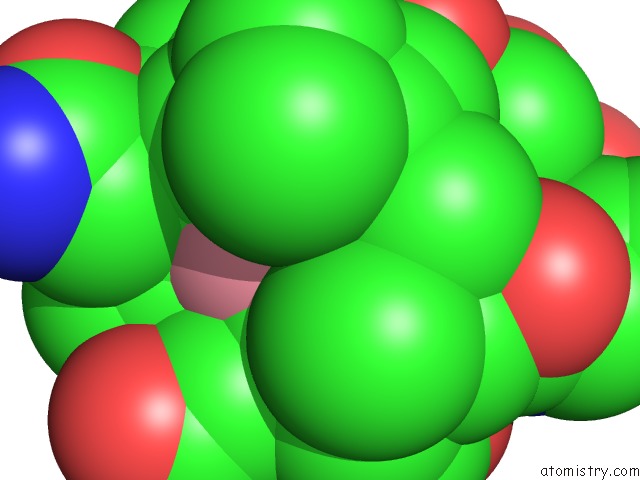
Mono view
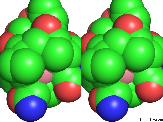
Stereo pair view
Mono view
Stereo pair view
A full contact list of Cobalt with other atoms in the Co binding
site number 1 of Crystal Structure of Diol Dehydratase-Cyanocobalamin Complex at 100K. within 5.0Å range:
|
Cobalt binding site 2 out of 2 in 1egm
Go back to
Cobalt binding site 2 out
of 2 in the Crystal Structure of Diol Dehydratase-Cyanocobalamin Complex at 100K.
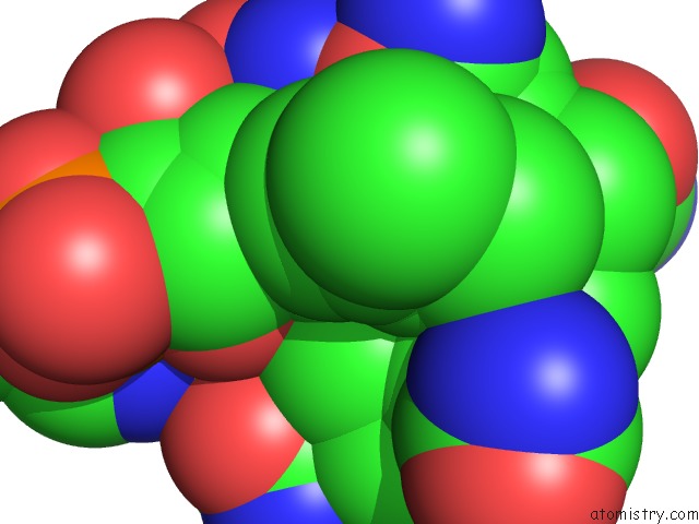
Mono view
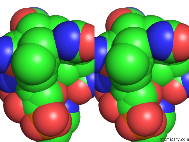
Stereo pair view
Mono view
Stereo pair view
A full contact list of Cobalt with other atoms in the Co binding
site number 2 of Crystal Structure of Diol Dehydratase-Cyanocobalamin Complex at 100K. within 5.0Å range:
|
Reference:
J.Masuda,
N.Shibata,
Y.Morimoto,
T.Toraya,
N.Yasuoka.
How A Protein Generates A Catalytic Radical From Coenzyme B(12): X-Ray Structure of A Diol-Dehydratase-Adeninylpentylcobalamin Complex. Structure Fold.Des. V. 8 775 2000.
ISSN: ISSN 0969-2126
PubMed: 10903944
DOI: 10.1016/S0969-2126(00)00164-7
Page generated: Tue Jul 30 14:13:28 2024
ISSN: ISSN 0969-2126
PubMed: 10903944
DOI: 10.1016/S0969-2126(00)00164-7
Last articles
Zn in 9J0NZn in 9J0O
Zn in 9J0P
Zn in 9FJX
Zn in 9EKB
Zn in 9C0F
Zn in 9CAH
Zn in 9CH0
Zn in 9CH3
Zn in 9CH1