Cobalt in PDB 1gqk: Structure of Pseudomonas Cellulosa Alpha-D-Glucuronidase Complexed with Glucuronic Acid
Enzymatic activity of Structure of Pseudomonas Cellulosa Alpha-D-Glucuronidase Complexed with Glucuronic Acid
All present enzymatic activity of Structure of Pseudomonas Cellulosa Alpha-D-Glucuronidase Complexed with Glucuronic Acid:
3.2.1.139;
Protein crystallography data
The structure of Structure of Pseudomonas Cellulosa Alpha-D-Glucuronidase Complexed with Glucuronic Acid, PDB code: 1gqk
was solved by
D.Nurizzo,
T.Nagy,
H.J.Gilbert,
G.J.Davies,
with X-Ray Crystallography technique. A brief refinement statistics is given in the table below:
Resolution Low / High (Å)
|
19.73 /
1.90
|
Space group
|
P 1
|
Cell size a, b, c (Å), α, β, γ (°)
|
69.707,
74.879,
87.481,
115.34,
93.06,
109.20
|
R / Rfree (%)
|
15.1 /
18.6
|
Cobalt Binding Sites:
The binding sites of Cobalt atom in the Structure of Pseudomonas Cellulosa Alpha-D-Glucuronidase Complexed with Glucuronic Acid
(pdb code 1gqk). This binding sites where shown within
5.0 Angstroms radius around Cobalt atom.
In total 8 binding sites of Cobalt where determined in the
Structure of Pseudomonas Cellulosa Alpha-D-Glucuronidase Complexed with Glucuronic Acid, PDB code: 1gqk:
Jump to Cobalt binding site number:
1;
2;
3;
4;
5;
6;
7;
8;
Cobalt binding site 1 out
of 8 in 1gqk
Go back to
Cobalt Binding Sites List in 1gqk
Cobalt binding site 1 out
of 8 in the Structure of Pseudomonas Cellulosa Alpha-D-Glucuronidase Complexed with Glucuronic Acid
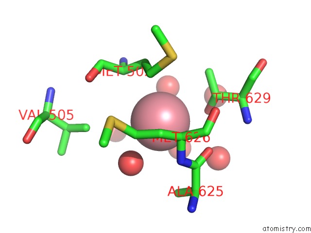
 Mono view
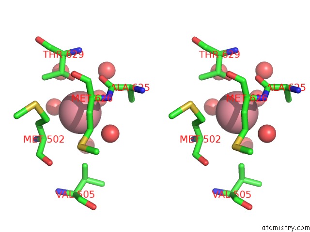
 Stereo pair view
|
A full contact list of Cobalt with other atoms in the Co binding
site number 1 of Structure of Pseudomonas Cellulosa Alpha-D-Glucuronidase Complexed with Glucuronic Acid within 5.0Å range:
probe
|
atom
|
residue
|
distance (Å)
|
B
|
Occ
|
A:Co1720
b:22.2
occ:0.50
|
OG1
|
A:THR629
|
3.0
|
14.0
|
1.0
|
O
|
A:HOH2546
|
3.1
|
20.6
|
1.0
|
O
|
A:HOH2439
|
3.4
|
23.2
|
1.0
|
O
|
A:HOH2649
|
3.5
|
17.6
|
1.0
|
O
|
A:ALA625
|
3.6
|
13.9
|
1.0
|
CB
|
A:THR629
|
3.7
|
15.1
|
1.0
|
CA
|
A:MET626
|
3.8
|
14.6
|
1.0
|
CA
|
A:MET502
|
3.8
|
16.0
|
1.0
|
CB
|
A:MET502
|
3.8
|
15.6
|
1.0
|
C
|
A:ALA625
|
3.8
|
13.2
|
1.0
|
N
|
A:MET626
|
3.9
|
13.9
|
1.0
|
CG
|
A:MET502
|
3.9
|
15.8
|
1.0
|
CG
|
A:MET626
|
4.1
|
17.4
|
1.0
|
CG2
|
A:THR629
|
4.2
|
14.8
|
1.0
|
N
|
A:MET502
|
4.4
|
16.2
|
1.0
|
CB
|
A:ALA625
|
4.5
|
12.2
|
1.0
|
O
|
A:HOH2547
|
4.5
|
19.4
|
1.0
|
CB
|
A:MET626
|
4.5
|
14.4
|
1.0
|
CG1
|
A:VAL505
|
4.5
|
18.2
|
1.0
|
CA
|
A:ALA625
|
4.8
|
13.4
|
1.0
|
O
|
A:HOH2170
|
4.8
|
39.1
|
1.0
|
O
|
A:HOH2554
|
4.8
|
24.1
|
1.0
|
C
|
A:MET626
|
4.9
|
14.1
|
1.0
|
|
Cobalt binding site 2 out
of 8 in 1gqk
Go back to
Cobalt Binding Sites List in 1gqk
Cobalt binding site 2 out
of 8 in the Structure of Pseudomonas Cellulosa Alpha-D-Glucuronidase Complexed with Glucuronic Acid
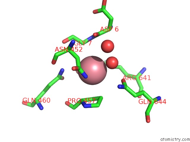
 Mono view
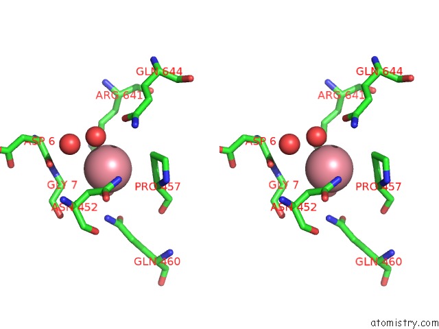
 Stereo pair view
|
A full contact list of Cobalt with other atoms in the Co binding
site number 2 of Structure of Pseudomonas Cellulosa Alpha-D-Glucuronidase Complexed with Glucuronic Acid within 5.0Å range:
probe
|
atom
|
residue
|
distance (Å)
|
B
|
Occ
|
A:Co1721
b:17.6
occ:0.50
|
O
|
A:HOH2572
|
2.9
|
20.5
|
1.0
|
O
|
A:HOH2412
|
3.0
|
13.9
|
1.0
|
ND2
|
A:ASN452
|
3.3
|
16.3
|
1.0
|
CB
|
A:ASN452
|
3.5
|
13.5
|
1.0
|
CB
|
A:PRO457
|
3.6
|
13.4
|
1.0
|
CG
|
A:PRO457
|
3.6
|
12.4
|
1.0
|
CA
|
A:GLY7
|
3.7
|
16.3
|
1.0
|
NE2
|
A:GLN460
|
3.7
|
20.4
|
1.0
|
CD
|
A:ARG641
|
3.8
|
13.1
|
1.0
|
CG
|
A:ASN452
|
3.9
|
15.1
|
1.0
|
CA
|
A:PRO457
|
4.0
|
13.4
|
1.0
|
N
|
A:GLY7
|
4.2
|
17.6
|
1.0
|
CG
|
A:ARG641
|
4.2
|
12.0
|
1.0
|
OE1
|
A:GLN644
|
4.3
|
17.5
|
1.0
|
CD
|
A:GLN460
|
4.4
|
15.1
|
1.0
|
OE1
|
A:GLN460
|
4.5
|
13.9
|
1.0
|
O
|
A:ASP6
|
4.5
|
18.4
|
1.0
|
C
|
A:ASP6
|
4.5
|
18.6
|
1.0
|
CD
|
A:PRO457
|
4.6
|
11.8
|
1.0
|
N
|
A:PRO457
|
4.7
|
13.6
|
1.0
|
O
|
A:HOH2010
|
4.7
|
24.8
|
1.0
|
C
|
A:GLY7
|
4.8
|
15.8
|
1.0
|
CA
|
A:ASN452
|
4.9
|
13.5
|
1.0
|
O
|
A:GLY7
|
5.0
|
15.7
|
1.0
|
|
Cobalt binding site 3 out
of 8 in 1gqk
Go back to
Cobalt Binding Sites List in 1gqk
Cobalt binding site 3 out
of 8 in the Structure of Pseudomonas Cellulosa Alpha-D-Glucuronidase Complexed with Glucuronic Acid
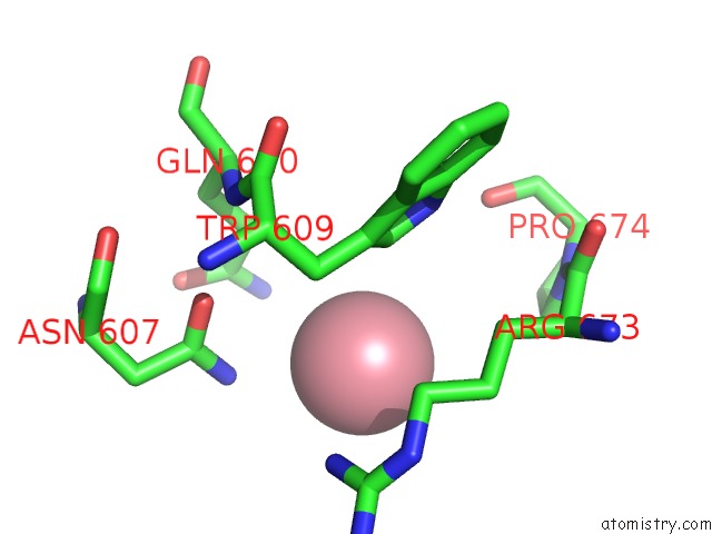
 Mono view
 Stereo pair view
|
A full contact list of Cobalt with other atoms in the Co binding
site number 3 of Structure of Pseudomonas Cellulosa Alpha-D-Glucuronidase Complexed with Glucuronic Acid within 5.0Å range:
probe
|
atom
|
residue
|
distance (Å)
|
B
|
Occ
|
A:Co1722
b:19.4
occ:0.50
|
NH1
|
A:ARG673
|
3.4
|
22.9
|
1.0
|
ND2
|
A:ASN607
|
3.4
|
17.8
|
1.0
|
CD
|
A:ARG673
|
3.6
|
24.3
|
1.0
|
CD1
|
A:TRP609
|
3.7
|
17.7
|
1.0
|
CG
|
A:ARG673
|
4.2
|
24.0
|
1.0
|
NE2
|
A:GLN610
|
4.2
|
24.3
|
1.0
|
CB
|
A:ARG673
|
4.2
|
23.6
|
1.0
|
CZ
|
A:ARG673
|
4.3
|
22.5
|
1.0
|
CG
|
A:ASN607
|
4.3
|
18.6
|
1.0
|
NE
|
A:ARG673
|
4.4
|
23.3
|
1.0
|
OD1
|
A:ASN607
|
4.4
|
14.5
|
1.0
|
CG
|
A:TRP609
|
4.4
|
16.0
|
1.0
|
CG
|
A:GLN610
|
4.5
|
21.0
|
1.0
|
CB
|
A:TRP609
|
4.5
|
16.4
|
1.0
|
CD
|
A:GLN610
|
4.6
|
23.4
|
1.0
|
NE1
|
A:TRP609
|
4.6
|
17.5
|
1.0
|
CD
|
A:PRO674
|
5.0
|
25.9
|
1.0
|
|
Cobalt binding site 4 out
of 8 in 1gqk
Go back to
Cobalt Binding Sites List in 1gqk
Cobalt binding site 4 out
of 8 in the Structure of Pseudomonas Cellulosa Alpha-D-Glucuronidase Complexed with Glucuronic Acid
 Mono view
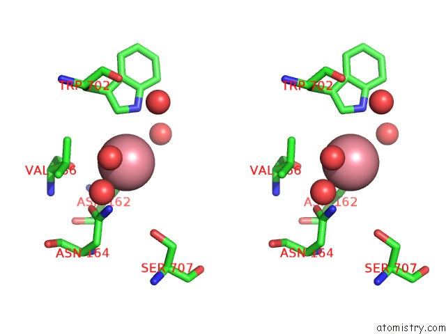
 Stereo pair view
|
A full contact list of Cobalt with other atoms in the Co binding
site number 4 of Structure of Pseudomonas Cellulosa Alpha-D-Glucuronidase Complexed with Glucuronic Acid within 5.0Å range:
probe
|
atom
|
residue
|
distance (Å)
|
B
|
Occ
|
A:Co1723
b:28.1
occ:0.50
|
O
|
A:HOH2650
|
2.7
|
22.5
|
1.0
|
ND2
|
A:ASN162
|
3.2
|
15.8
|
1.0
|
ND2
|
A:ASN164
|
3.3
|
17.8
|
1.0
|
CD1
|
A:TRP702
|
3.6
|
19.6
|
1.0
|
CB
|
A:VAL166
|
3.8
|
15.3
|
1.0
|
OD1
|
A:ASN164
|
3.8
|
14.0
|
1.0
|
NE1
|
A:TRP702
|
3.9
|
18.2
|
1.0
|
CG
|
A:ASN164
|
3.9
|
14.7
|
1.0
|
CG1
|
A:VAL166
|
3.9
|
16.6
|
1.0
|
CG
|
A:ASN162
|
4.2
|
16.3
|
1.0
|
CB
|
A:ASN162
|
4.4
|
14.9
|
1.0
|
O
|
A:HOH2628
|
4.4
|
41.5
|
1.0
|
CG2
|
A:VAL166
|
4.5
|
16.6
|
1.0
|
OG
|
B:SER707
|
4.8
|
18.8
|
1.0
|
CG
|
A:TRP702
|
4.8
|
17.3
|
1.0
|
O
|
A:VAL166
|
4.8
|
14.2
|
1.0
|
O
|
A:HOH2157
|
4.9
|
25.0
|
1.0
|
O
|
B:HOH2609
|
5.0
|
19.7
|
1.0
|
CA
|
A:VAL166
|
5.0
|
14.6
|
1.0
|
|
Cobalt binding site 5 out
of 8 in 1gqk
Go back to
Cobalt Binding Sites List in 1gqk
Cobalt binding site 5 out
of 8 in the Structure of Pseudomonas Cellulosa Alpha-D-Glucuronidase Complexed with Glucuronic Acid
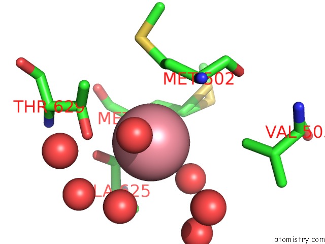
 Mono view
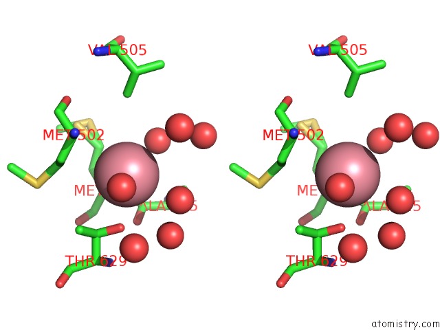
 Stereo pair view
|
A full contact list of Cobalt with other atoms in the Co binding
site number 5 of Structure of Pseudomonas Cellulosa Alpha-D-Glucuronidase Complexed with Glucuronic Acid within 5.0Å range:
probe
|
atom
|
residue
|
distance (Å)
|
B
|
Occ
|
B:Co1720
b:22.7
occ:0.50
|
OG1
|
B:THR629
|
3.0
|
13.7
|
1.0
|
O
|
B:HOH2528
|
3.2
|
20.1
|
1.0
|
O
|
B:HOH2634
|
3.4
|
16.5
|
1.0
|
O
|
B:HOH2428
|
3.4
|
23.6
|
1.0
|
CB
|
B:THR629
|
3.7
|
14.5
|
1.0
|
O
|
B:ALA625
|
3.7
|
14.5
|
1.0
|
CB
|
B:MET502
|
3.7
|
15.6
|
1.0
|
CA
|
B:MET626
|
3.8
|
14.7
|
1.0
|
CA
|
B:MET502
|
3.8
|
16.0
|
1.0
|
CG
|
B:MET502
|
3.8
|
18.1
|
1.0
|
C
|
B:ALA625
|
3.8
|
14.7
|
1.0
|
N
|
B:MET626
|
3.9
|
14.6
|
1.0
|
CG
|
B:MET626
|
4.1
|
13.0
|
0.5
|
CG
|
B:MET626
|
4.1
|
15.6
|
0.5
|
CG2
|
B:THR629
|
4.1
|
16.2
|
1.0
|
O
|
B:HOH2530
|
4.2
|
19.2
|
1.0
|
N
|
B:MET502
|
4.4
|
15.4
|
1.0
|
CB
|
B:MET626
|
4.5
|
15.3
|
1.0
|
CB
|
B:ALA625
|
4.5
|
14.9
|
1.0
|
CG1
|
B:VAL505
|
4.6
|
19.2
|
1.0
|
O
|
B:HOH2162
|
4.7
|
34.9
|
1.0
|
O
|
B:HOH2537
|
4.8
|
25.0
|
1.0
|
CA
|
B:ALA625
|
4.8
|
14.8
|
1.0
|
C
|
B:MET626
|
4.9
|
15.7
|
1.0
|
O
|
B:HOH2203
|
5.0
|
19.8
|
1.0
|
|
Cobalt binding site 6 out
of 8 in 1gqk
Go back to
Cobalt Binding Sites List in 1gqk
Cobalt binding site 6 out
of 8 in the Structure of Pseudomonas Cellulosa Alpha-D-Glucuronidase Complexed with Glucuronic Acid
 Mono view
 Stereo pair view
|
A full contact list of Cobalt with other atoms in the Co binding
site number 6 of Structure of Pseudomonas Cellulosa Alpha-D-Glucuronidase Complexed with Glucuronic Acid within 5.0Å range:
probe
|
atom
|
residue
|
distance (Å)
|
B
|
Occ
|
B:Co1721
b:18.1
occ:0.50
|
O
|
B:HOH2402
|
3.0
|
13.3
|
1.0
|
O
|
B:HOH2556
|
3.0
|
18.0
|
1.0
|
ND2
|
B:ASN452
|
3.3
|
14.9
|
1.0
|
CB
|
B:ASN452
|
3.5
|
14.6
|
1.0
|
NE2
|
B:GLN460
|
3.6
|
19.2
|
1.0
|
CA
|
B:GLY7
|
3.6
|
17.5
|
1.0
|
CB
|
B:PRO457
|
3.7
|
14.0
|
1.0
|
CG
|
B:PRO457
|
3.7
|
13.5
|
1.0
|
CD
|
B:ARG641
|
3.9
|
16.1
|
1.0
|
CG
|
B:ASN452
|
3.9
|
13.6
|
1.0
|
CA
|
B:PRO457
|
4.0
|
13.4
|
1.0
|
N
|
B:GLY7
|
4.1
|
18.0
|
1.0
|
CG
|
B:ARG641
|
4.3
|
13.6
|
1.0
|
CD
|
B:GLN460
|
4.3
|
18.4
|
1.0
|
OE1
|
B:GLN460
|
4.4
|
17.8
|
1.0
|
O
|
B:ASP6
|
4.4
|
19.4
|
1.0
|
C
|
B:ASP6
|
4.4
|
19.9
|
1.0
|
OE1
|
B:GLN644
|
4.5
|
17.6
|
1.0
|
O
|
B:HOH2010
|
4.7
|
25.8
|
1.0
|
C
|
B:GLY7
|
4.7
|
16.5
|
1.0
|
CD
|
B:PRO457
|
4.8
|
12.3
|
1.0
|
N
|
B:PRO457
|
4.8
|
12.9
|
1.0
|
CA
|
B:ASN452
|
4.8
|
14.2
|
1.0
|
O
|
B:GLY7
|
4.9
|
16.6
|
1.0
|
|
Cobalt binding site 7 out
of 8 in 1gqk
Go back to
Cobalt Binding Sites List in 1gqk
Cobalt binding site 7 out
of 8 in the Structure of Pseudomonas Cellulosa Alpha-D-Glucuronidase Complexed with Glucuronic Acid
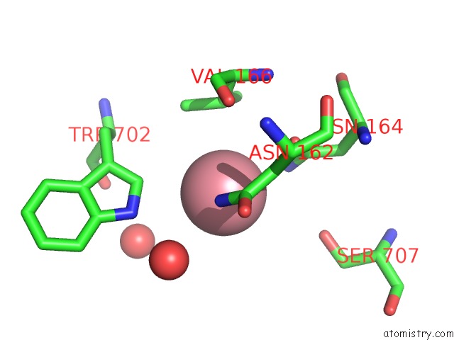
 Mono view
 Stereo pair view
|
A full contact list of Cobalt with other atoms in the Co binding
site number 7 of Structure of Pseudomonas Cellulosa Alpha-D-Glucuronidase Complexed with Glucuronic Acid within 5.0Å range:
probe
|
atom
|
residue
|
distance (Å)
|
B
|
Occ
|
B:Co1722
b:28.4
occ:0.50
|
O
|
B:HOH2635
|
2.7
|
24.0
|
1.0
|
ND2
|
B:ASN162
|
3.1
|
18.0
|
1.0
|
ND2
|
B:ASN164
|
3.3
|
18.8
|
1.0
|
OD1
|
B:ASN164
|
3.7
|
15.2
|
1.0
|
CD1
|
B:TRP702
|
3.7
|
17.6
|
1.0
|
CB
|
B:VAL166
|
3.8
|
13.8
|
1.0
|
CG
|
B:ASN164
|
3.9
|
16.3
|
1.0
|
CG1
|
B:VAL166
|
4.0
|
16.3
|
1.0
|
CG
|
B:ASN162
|
4.1
|
17.0
|
1.0
|
NE1
|
B:TRP702
|
4.1
|
18.0
|
1.0
|
CB
|
B:ASN162
|
4.2
|
13.9
|
1.0
|
CG2
|
B:VAL166
|
4.4
|
16.9
|
1.0
|
O
|
B:HOH2611
|
4.6
|
36.6
|
1.0
|
O
|
B:VAL166
|
4.7
|
13.8
|
1.0
|
OG
|
A:SER707
|
4.8
|
20.5
|
1.0
|
CA
|
B:VAL166
|
4.9
|
14.4
|
1.0
|
CG
|
B:TRP702
|
4.9
|
17.3
|
1.0
|
O
|
B:HOH2152
|
5.0
|
26.6
|
1.0
|
|
Cobalt binding site 8 out
of 8 in 1gqk
Go back to
Cobalt Binding Sites List in 1gqk
Cobalt binding site 8 out
of 8 in the Structure of Pseudomonas Cellulosa Alpha-D-Glucuronidase Complexed with Glucuronic Acid
 Mono view
 Stereo pair view
|
A full contact list of Cobalt with other atoms in the Co binding
site number 8 of Structure of Pseudomonas Cellulosa Alpha-D-Glucuronidase Complexed with Glucuronic Acid within 5.0Å range:
probe
|
atom
|
residue
|
distance (Å)
|
B
|
Occ
|
B:Co1723
b:22.5
occ:0.50
|
NH1
|
B:ARG673
|
3.3
|
26.0
|
1.0
|
ND2
|
B:ASN607
|
3.4
|
18.9
|
1.0
|
CD1
|
B:TRP609
|
3.7
|
18.2
|
1.0
|
CD
|
B:ARG673
|
3.7
|
25.4
|
1.0
|
OD1
|
B:ASN607
|
4.2
|
15.7
|
1.0
|
NE2
|
B:GLN610
|
4.2
|
20.7
|
1.0
|
CG
|
B:ASN607
|
4.2
|
17.0
|
1.0
|
CG
|
B:ARG673
|
4.3
|
26.2
|
1.0
|
CZ
|
B:ARG673
|
4.3
|
23.8
|
1.0
|
CB
|
B:ARG673
|
4.3
|
26.1
|
1.0
|
NE
|
B:ARG673
|
4.5
|
24.4
|
1.0
|
CG
|
B:TRP609
|
4.5
|
16.6
|
1.0
|
CG
|
B:GLN610
|
4.5
|
18.8
|
1.0
|
CD
|
B:GLN610
|
4.6
|
21.4
|
1.0
|
CB
|
B:TRP609
|
4.6
|
17.1
|
1.0
|
NE1
|
B:TRP609
|
4.6
|
17.9
|
1.0
|
CD
|
B:PRO674
|
4.9
|
27.7
|
1.0
|
|
Reference:
D.Nurizzo,
T.Nagy,
H.J.Gilbert,
G.J.Davies.
The Structural Basis For Catalysis and Specificity of the Pseudomonas Cellulosa Alpha-Glucuronidase, GLCA67A Structure V. 10 547 2002.
ISSN: ISSN 0969-2126
PubMed: 11937059
DOI: 10.1016/S0969-2126(02)00742-6
Page generated: Tue Jul 30 14:17:15 2024
|